

| << Chapter < Page | Chapter >> Page > |
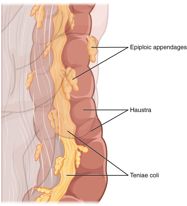
Three features are unique to the large intestine: teniae coli, haustra, and epiploic appendages ( [link] ). The teniae coli are three bands of smooth muscle that make up the longitudinal muscle layer of the muscularis of the large intestine, except at its terminal end. Tonic contractions of the teniae coli bunch up the colon into a succession of pouches called haustra (singular = hostrum), which are responsible for the wrinkled appearance of the colon. Attached to the teniae coli are small, fat-filled sacs of visceral peritoneum called epiploic appendages . The purpose of these is unknown. Although the rectum and anal canal have neither teniae coli nor haustra, they do have well-developed layers of muscularis that create the strong contractions needed for defecation.
Most bacteria that enter the alimentary canal are killed by lysozyme, HCl, or protein-digesting enzymes. However, trillions of bacteria live within the large intestine and are referred to as the bacterial flora . Most of the more than 700 species of these bacteria cause no harm as long as they stay in the gut lumen. In fact, many facilitate chemical digestion and absorption.
The residue of chyme that enters the large intestine contains few nutrients except water, which is reabsorbed as the residue lingers in the large intestine, typically for 12 to 24 hours. Thus, it may not surprise you that the large intestine can be completely removed without significantly affecting digestive functioning. For example, in severe cases of inflammatory bowel disease, the large intestine can be removed by a procedure known as a colectomy. Often, a new fecal pouch can be crafted from the small intestine and sutured to the anus, but if not, an ileostomy can be created by bringing the distal ileum through the abdominal wall, allowing the watery chyme to be collected in a bag-like adhesive appliance.
The small intestine absorbs about 90 percent of the water you ingest (either as liquid or within solid food). The large intestine absorbs most of the remaining water, a process that converts the liquid chyme residue into semisolid feces (“stool”). Feces is composed of undigested food residues, unabsorbed digested substances, millions of bacteria, old epithelial cells from the GI mucosa, inorganic salts, and enough water to let it pass smoothly out of the body. Of every 500 mL (17 ounces) of food residue that enters the cecum each day, about 150 mL (5 ounces) become feces.
Feces are eliminated through contractions of the rectal muscles. You help this process by a voluntary procedure called Valsalva’s maneuver , in which you increase intra-abdominal pressure by contracting your diaphragm and abdominal wall muscles, and closing your glottis.
If defecation is delayed for an extended time, additional water is absorbed, making the feces firmer and potentially leading to constipation. On the other hand, if the waste matter moves too quickly through the intestines, not enough water is absorbed, and diarrhea can result. This can be caused by the ingestion of foodborne pathogens. In general, diet, health, and stress determine the frequency of bowel movements. The number of bowel movements varies greatly between individuals, ranging from two or three per day to three or four per week.
The three main regions of the small intestine are the duodenum, the jejunum, and the ileum. The small intestine is where digestion is completed and virtually all absorption occurs. These two activities are facilitated by structural adaptations that increase the mucosal surface area by 600-fold, including circular folds, villi, and microvilli. There are around 200 million microvilli per square millimeter of small intestine, which contain brush border enzymes that complete the digestion of carbohydrates and proteins. Combined with pancreatic juice, intestinal juice provides the liquid medium needed to further digest and absorb substances from chyme. The small intestine is also the site of unique mechanical digestive movements. Segmentation moves the chyme back and forth, increasing mixing and opportunities for absorption. Migrating motility complexes propel the residual chyme toward the large intestine.
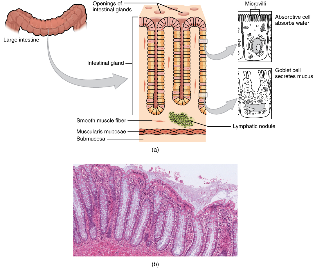
The main regions of the large intestine are the cecum, the colon, and the rectum. The large intestine absorbs water and forms feces, and is responsible for defecation. Bacterial flora break down additional carbohydrate residue, and synthesize certain vitamins. The mucosa of the large intestinal wall is generously endowed with goblet cells, which secrete mucus that eases the passage of feces. The entry of feces into the rectum activates the defecation reflex.
American Cancer Society (US). Cancer facts and figures: colorectal cancer: 2011–2013 [Internet]. c2013 [cited 2013 Apr 3]. Available from: (External Link) .
The Nutrition Source. Fiber and colon cancer: following the scientific trail [Internet]. Boston (MA): Harvard School of Public Health; c2012 [cited 2013 Apr 3]. Available from: (External Link) .
Centers for Disease Control and Prevention (US). Morbidity and mortality weekly report: notifiable diseases and mortality tables [Internet]. Atlanta (GA); [cited 2013 Apr 3]. Available from: (External Link) .
Notification Switch
Would you like to follow the 'Digestive system' conversation and receive update notifications?

|
|

|
|

|
|

|
|

|
|

|
|

|
|

|
|

|
|

|