

| << Chapter < Page | Chapter >> Page > |
The large intestine is the terminal part of the alimentary canal. The primary function of this organ is to finish absorption of nutrients and water, synthesize certain vitamins, form feces, and eliminate feces from the body.
The large intestine runs from the appendix to the anus. It frames the small intestine on three sides. Despite its being about one-half as long as the small intestine, it is called large because it is more than twice the diameter of the small intestine, about 3 inches. The large intestine is subdivided into four main regions: the cecum, the colon, the rectum, and the anus. The ileocecal valve, located at the opening between the ileum and the large intestine, controls the flow of chyme from the small intestine to the large intestine.
The first part of the large intestine is the cecum , a sac-like structure that is suspended inferior to the ileocecal valve. It is about 2.4 inches long, receives the contents of the ileum, and continues the absorption of water and salts. The appendix is a winding tube that attaches to the cecum. Although the 3-inch long appendix contains lymphoid tissue, suggesting an immune function, this organ is generally considered vestigial (no longer useful). However, at least one recent report suggests a survival advantage provided by the appendix: In illness, the appendix may serve as a bacterial reservoir to repopulate the bacteria after the illness.
The cecum blends seamlessly with the colon . Upon entering the colon, the food residue first travels up the ascending colon on the right side of the abdomen. At the inferior surface of the liver, the colon bends to form the right colic flexure (hepatic flexure) and becomes the transverse colon . Food residue passing through the transverse colon travels across to the left side of the abdomen, where the colon angles sharply immediately inferior to the spleen, at the left colic flexure (splenic flexure). From there, food residue passes through the descending colon , which runs down the left side of the abdominal wall. After entering the pelvis it becomes the s-shaped sigmoid colon .
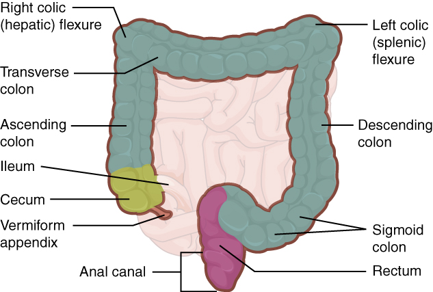
Food residue leaving the sigmoid colon enters the rectum in the pelvis. The final 8 inches of the alimentary canal, the rectum extends to the sacrum and coccyx. The rectum stores formed feces until the body is ready to expel the waste. Finally, food residue reaches the last part of the large intestine, the anal canal which opens to the exterior of the body at the anus. The anal canal includes two sphincters. The internal anal sphincter is made of smooth muscle, and its contractions are involuntary. The external anal sphincter is made of skeletal muscle, which is under voluntary control. Except when defecating, both usually remain closed.
There are several notable differences between the walls of the large and small intestines ( [link] ). For example, few enzyme-secreting cells are found in the wall of the large intestine, and there are no circular folds or villi. There is an increased number of mucus producing goblet cells . These goblet cells secrete mucus that eases the movement of feces and protects the intestine.
Notification Switch
Would you like to follow the 'Digestive system' conversation and receive update notifications?

|
|

|
|

|
|

|
|

|
|

|
|

|
|

|
|

|
|

|