<< Chapter < Page Chapter >> Page >

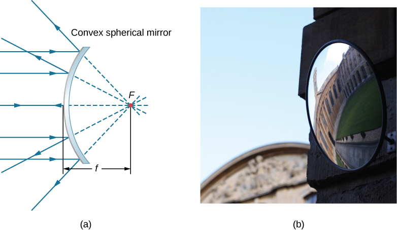
A convex spherical mirror also has a focal point, as shown in [link] . Incident rays parallel to the optical axis are reflected from the mirror and seem to originate from point F at focal length f behind the mirror. Thus, the focal point is virtual because no real rays actually pass through it; they only appear to originate from it.

Figure a shows the cross section of a convex mirror. Parallel rays reflect from it and diverge in different directions. The reflected rays are extended at the back by dotted lines and seem to originate from a single point behind the mirror. This point is labeled F. The distance from this point to the mirror is labeled f. Figure b shows the photograph of a convex mirror reflecting the image of a building. The image is curved and distorted.
(a) Rays reflected by a convex spherical mirror: Incident rays of light parallel to the optical axis are reflected from a convex spherical mirror and seem to originate from a well-defined focal point at focal distance f on the opposite side of the mirror. The focal point is virtual because no real rays pass through it. (b) Photograph of a virtual image formed by a convex mirror. (credit b: modification of work by Jenny Downing)

How does the focal length of a mirror relate to the mirror’s radius of curvature? [link] shows a single ray that is reflected by a spherical concave mirror. The incident ray is parallel to the optical axis. The point at which the reflected ray crosses the optical axis is the focal point. Note that all incident rays that are parallel to the optical axis are reflected through the focal point—we only show one ray for simplicity. We want to find how the focal length FP (denoted by f ) relates to the radius of curvature of the mirror, R , whose length is R = C F + F P . The law of reflection tells us that angles OXC and CXF are the same, and because the incident ray is parallel to the optical axis, angles OXC and XCP are also the same. Thus, triangle CXF is an isosceles triangle with C F = F X . If the angle θ is small (so that sin θ θ ; this is called the “small-angle approximation”), then F X F P or C F F P . Inserting this into the equation for the radius R , we get

R = C F + F P = F P + F P = 2 F P = 2 f
Figure shows the diagram of a concave mirror. An incident ray starting from point O hits the mirror at point X. The reflected ray passes through point F. A line CX bisects the angle formed by the incident and reflected rays. This line is labeled R. A line parallel to the incident ray passes through points C and F and hits the mirror at point P. The distance between points F and P is labeled f. Angle OXC, angle CXF and angle XCF are all labeled theta.
Reflection in a concave mirror. In the small-angle approximation, a ray that is parallel to the optical axis CP is reflected through the focal point F of the mirror.

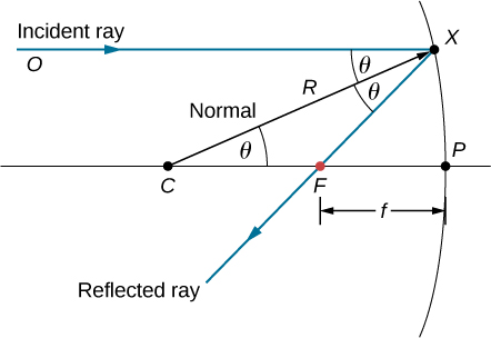
In other words, in the small-angle approximation, the focal length f of a concave spherical mirror is half of its radius of curvature, R :

f = R 2 .

In this chapter, we assume that the small-angle approximation    (also called the paraxial approximation) is always valid. In this approximation, all rays are paraxial rays, which means that they make a small angle with the optical axis and are at a distance much less than the radius of curvature from the optical axis. In this case, their angles θ of reflection are small angles, so sin θ tan θ θ .

Using ray tracing to locate images

To find the location of an image formed by a spherical mirror, we first use ray tracing , which is the technique of drawing rays and using the law of reflection to determine the reflected rays (later, for lenses, we use the law of refraction to determine refracted rays). Combined with some basic geometry, we can use ray tracing to find the focal point, the image location, and other information about how a mirror manipulates light. In fact, we already used ray tracing above to locate the focal point of spherical mirrors, or the image distance of flat mirrors. To locate the image of an object, you must locate at least two points of the image. Locating each point requires drawing at least two rays from a point on the object and constructing their reflected rays. The point at which the reflected rays intersect, either in real space or in virtual space, is where the corresponding point of the image is located. To make ray tracing easier, we concentrate on four “principal” rays whose reflections are easy to construct.

Questions & Answers

what is phylogeny
Odigie Reply
evolutionary history and relationship of an organism or group of organisms
AI-Robot
ok
Deng
what is biology
Hajah Reply
the study of living organisms and their interactions with one another and their environments
AI-Robot
what is biology
Victoria Reply
HOW CAN MAN ORGAN FUNCTION
Alfred Reply
the diagram of the digestive system
Assiatu Reply
allimentary cannel
Ogenrwot
How does twins formed
William Reply
They formed in two ways first when one sperm and one egg are splited by mitosis or two sperm and two eggs join together
Oluwatobi
what is genetics
Josephine Reply
Genetics is the study of heredity
Misack
how does twins formed?
Misack
What is manual
Hassan Reply
discuss biological phenomenon and provide pieces of evidence to show that it was responsible for the formation of eukaryotic organelles
Joseph Reply
what is biology
Yousuf Reply
the study of living organisms and their interactions with one another and their environment.
Wine
discuss the biological phenomenon and provide pieces of evidence to show that it was responsible for the formation of eukaryotic organelles in an essay form
Joseph Reply
what is the blood cells
Shaker Reply
list any five characteristics of the blood cells
Shaker
lack electricity and its more savely than electronic microscope because its naturally by using of light
Abdullahi Reply
advantage of electronic microscope is easily and clearly while disadvantage is dangerous because its electronic. advantage of light microscope is savely and naturally by sun while disadvantage is not easily,means its not sharp and not clear
Abdullahi
cell theory state that every organisms composed of one or more cell,cell is the basic unit of life
Abdullahi
is like gone fail us
DENG
cells is the basic structure and functions of all living things
Ramadan
What is classification
ISCONT Reply
is organisms that are similar into groups called tara
Yamosa
in what situation (s) would be the use of a scanning electron microscope be ideal and why?
Kenna Reply
A scanning electron microscope (SEM) is ideal for situations requiring high-resolution imaging of surfaces. It is commonly used in materials science, biology, and geology to examine the topography and composition of samples at a nanoscale level. SEM is particularly useful for studying fine details,
Hilary
Got questions? Join the online conversation and get instant answers!
Jobilize.com Reply

Get Jobilize Job Search Mobile App in your pocket Now!

Get it on Google Play Download on the App Store Now


Source:  OpenStax, University physics volume 3. OpenStax CNX. Nov 04, 2016 Download for free at http://cnx.org/content/col12067/1.4
Google Play and the Google Play logo are trademarks of Google Inc.

Notification Switch

Would you like to follow the 'University physics volume 3' conversation and receive update notifications?

Ask