<< Chapter < Page Chapter >> Page >
By the end of this section, you will be able to:
  • Use ray diagrams to locate and describe the image formed by a lens
  • Employ the thin-lens equation to describe and locate the image formed by a lens

Lenses are found in a huge array of optical instruments, ranging from a simple magnifying glass to a camera’s zoom lens to the eye itself. In this section, we use the Snell’s law to explore the properties of lenses and how they form images.

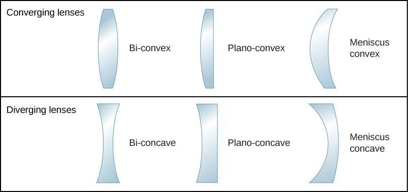
The word “lens” derives from the Latin word for a lentil bean, the shape of which is similar to a convex lens. However, not all lenses have the same shape. [link] shows a variety of different lens shapes. The vocabulary used to describe lenses is the same as that used for spherical mirrors: The axis of symmetry of a lens is called the optical axis, where this axis intersects the lens surface is called the vertex of the lens, and so forth.

Figure shows three converging lenses and three diverging lenses. The converging lenses are: bi-convex, with two convex surfaces, plano-convex, with one convex and once flat surface and meniscus convex, with one convex and one concave surface, the convex having a smaller radius of curvature. The diverging lenses are: bi-concave, with two concave surfaces, plano-concave, with one concave and once flat surface and meniscus concave, with one concave and one convex surface, the concave having a smaller radius of curvature.
Various types of lenses: Note that a converging lens has a thicker “waist,” whereas a diverging lens has a thinner waist.

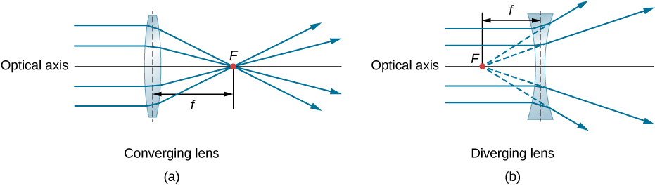
A convex or converging lens is shaped so that all light rays that enter it parallel to its optical axis intersect (or focus) at a single point on the optical axis on the opposite side of the lens, as shown in part (a) of [link] . Likewise, a concave or diverging lens is shaped so that all rays that enter it parallel to its optical axis diverge, as shown in part (b). To understand more precisely how a lens manipulates light, look closely at the top ray that goes through the converging lens in part (a). Because the index of refraction of the lens is greater than that of air, Snell’s law tells us that the ray is bent toward the perpendicular to the interface as it enters the lens. Likewise, when the ray exits the lens, it is bent away from the perpendicular. The same reasoning applies to the diverging lenses, as shown in part (b). The overall effect is that light rays are bent toward the optical axis for a converging lens and away from the optical axis for diverging lenses. For a converging lens, the point at which the rays cross is the focal point F of the lens. For a diverging lens, the point from which the rays appear to originate is the (virtual) focal point. The distance from the center of the lens to its focal point is the focal length f of the lens.

Figure a shows rays parallel to the optical axis striking a bi-convex lens and converging on the other side at point F. Figure b shows rays parallel to the optical axis striking a bi-concave lens and diverging on the other side. The diverging rays are extended at the back and seem to originate from point F in front of the lens. In both figures the distance from the center of the lens to point F is labeled f.
Rays of light entering (a) a converging lens and (b) a diverging lens, parallel to its axis, converge at its focal point F . The distance from the center of the lens to the focal point is the lens’s focal length f . Note that the light rays are bent upon entering and exiting the lens, with the overall effect being to bend the rays toward the optical axis.

A lens is considered to be thin if its thickness t is much less than the radii of curvature of both surfaces, as shown in [link] . In this case, the rays may be considered to bend once at the center of the lens. For the case drawn in the figure, light ray 1 is parallel to the optical axis, so the outgoing ray is bent once at the center of the lens and goes through the focal point. Another important characteristic of thin lenses is that light rays that pass through the center of the lens are undeviated, as shown by light ray 2.

Questions & Answers

what is biology
Hajah Reply
the study of living organisms and their interactions with one another and their environments
AI-Robot
what is biology
Victoria Reply
HOW CAN MAN ORGAN FUNCTION
Alfred Reply
the diagram of the digestive system
Assiatu Reply
allimentary cannel
Ogenrwot
How does twins formed
William Reply
They formed in two ways first when one sperm and one egg are splited by mitosis or two sperm and two eggs join together
Oluwatobi
what is genetics
Josephine Reply
Genetics is the study of heredity
Misack
how does twins formed?
Misack
What is manual
Hassan Reply
discuss biological phenomenon and provide pieces of evidence to show that it was responsible for the formation of eukaryotic organelles
Joseph Reply
what is biology
Yousuf Reply
the study of living organisms and their interactions with one another and their environment.
Wine
discuss the biological phenomenon and provide pieces of evidence to show that it was responsible for the formation of eukaryotic organelles in an essay form
Joseph Reply
what is the blood cells
Shaker Reply
list any five characteristics of the blood cells
Shaker
lack electricity and its more savely than electronic microscope because its naturally by using of light
Abdullahi Reply
advantage of electronic microscope is easily and clearly while disadvantage is dangerous because its electronic. advantage of light microscope is savely and naturally by sun while disadvantage is not easily,means its not sharp and not clear
Abdullahi
cell theory state that every organisms composed of one or more cell,cell is the basic unit of life
Abdullahi
is like gone fail us
DENG
cells is the basic structure and functions of all living things
Ramadan
What is classification
ISCONT Reply
is organisms that are similar into groups called tara
Yamosa
in what situation (s) would be the use of a scanning electron microscope be ideal and why?
Kenna Reply
A scanning electron microscope (SEM) is ideal for situations requiring high-resolution imaging of surfaces. It is commonly used in materials science, biology, and geology to examine the topography and composition of samples at a nanoscale level. SEM is particularly useful for studying fine details,
Hilary
cell is the building block of life.
Condoleezza Reply
Got questions? Join the online conversation and get instant answers!
Jobilize.com Reply
Practice Key Terms 5

Get Jobilize Job Search Mobile App in your pocket Now!

Get it on Google Play Download on the App Store Now


Source:  OpenStax, University physics volume 3. OpenStax CNX. Nov 04, 2016 Download for free at http://cnx.org/content/col12067/1.4
Google Play and the Google Play logo are trademarks of Google Inc.

Notification Switch

Would you like to follow the 'University physics volume 3' conversation and receive update notifications?

Ask