<< Chapter < Page Chapter >> Page >

Bone markings

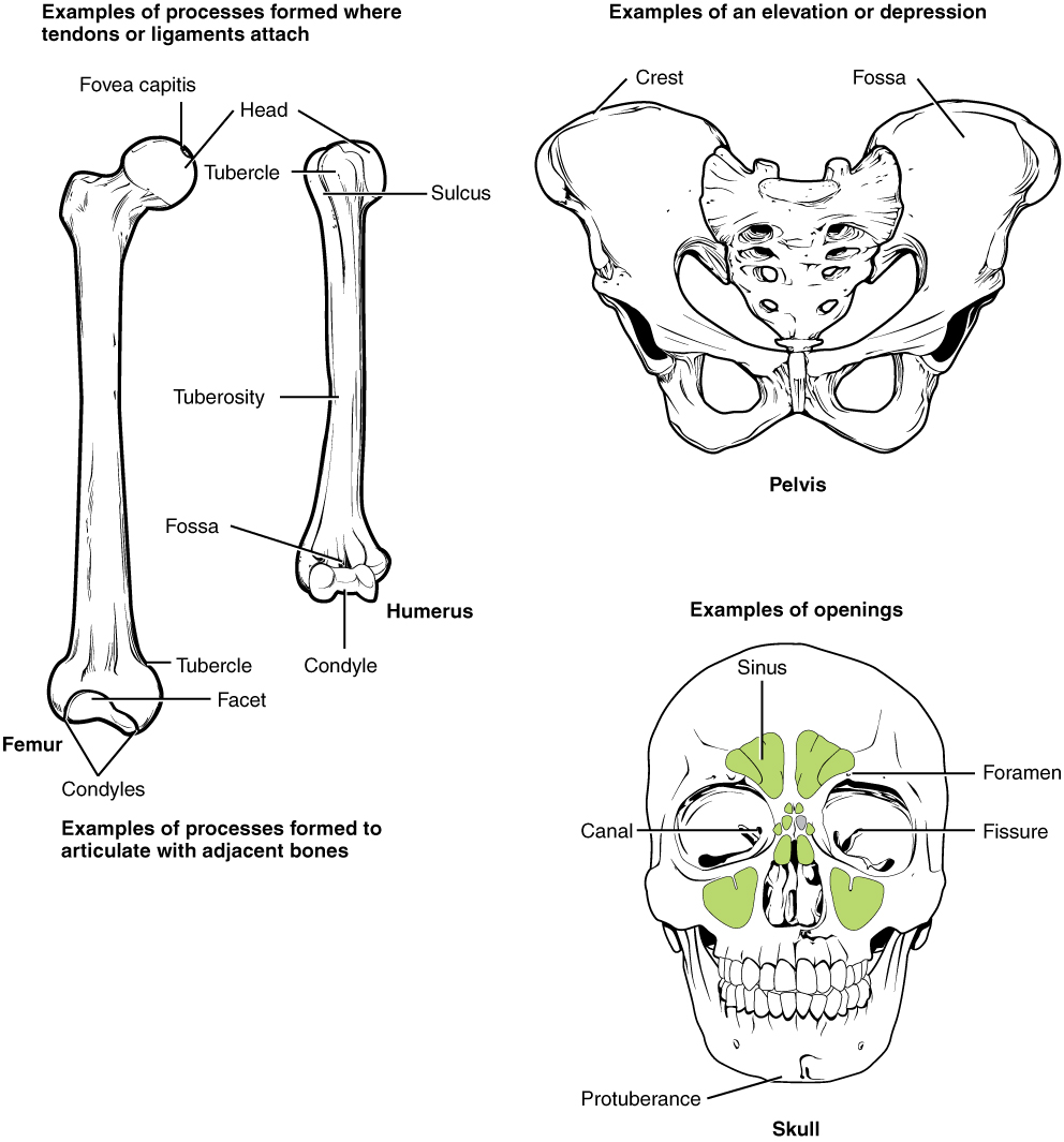
The surface features of bones vary considerably, depending on the function and location in the body. [link] describes the bone markings, which are illustrated in ( [link] ). There are three general classes of bone markings: (1) articulations, (2) projections, and (3) holes. As the name implies, an articulation    is where two bone surfaces come together (articulus = “joint”). These surfaces tend to conform to one another, such as one being rounded and the other cupped, to facilitate the function of the articulation. A projection    is an area of a bone that projects above the surface of the bone. These are the attachment points for tendons and ligaments. In general, their size and shape is an indication of the forces exerted through the attachment to the bone. A hole    is an opening or groove in the bone that allows blood vessels and nerves to enter the bone. As with the other markings, their size and shape reflect the size of the vessels and nerves that penetrate the bone at these points.

Bone Markings
Marking Description Example
Articulations Where two bones meet Knee joint
Head Prominent rounded surface Head of femur
Facet Flat surface Vertebrae
Condyle Rounded surface Occipital condyles
Projections Raised markings Spinous process of the vertebrae
Protuberance Protruding Chin
Process Prominence feature Transverse process of vertebra
Spine Sharp process Ischial spine
Tubercle Small, rounded process Tubercle of humerus
Tuberosity Rough surface Deltoid tuberosity
Line Slight, elongated ridge Temporal lines of the parietal bones
Crest Ridge Iliac crest
Holes Holes and depressions Foramen (holes through which blood vessels can pass through)
Fossa Elongated basin Mandibular fossa
Fovea Small pit Fovea capitis on the head of the femur
Sulcus Groove Sigmoid sulcus of the temporal bones
Canal Passage in bone Auditory canal
Fissure Slit through bone Auricular fissure
Foramen Hole through bone Foramen magnum in the occipital bone
Meatus Opening into canal External auditory meatus
Sinus Air-filled space in bone Nasal sinus

Bone features

This illustration contains three diagrams. The left diagram is titled examples of processes formed where tendons or ligaments attach. The image shows an anterior view of the femur and an anterior view of the humerus. For the femur, the distal epiphysis contains a smaller lateral bulge and a larger medial bulge. These are examples of condyles. The inner halves of the two condyles as well as the groove between them compose a facet. An oval-shaped ridge on the medial surface of the distal metaphysis is an example of a tubercle. On the proximal epiphysis of the femur, the large knob that attaches to the hip socket is an example of a head. The tip of the head contains a small depression, an example of a fovea called the fovea capitis. On the humerus, the distal epiphysis contains a central depression that is an example of a fossa. Two condyles are located on the right and left sides of the fossa. The diaphysis of the humerus contains a small ridge running up the shaft that is an example of a tuberosity. The proximal epiphysis of the humerus contains a lateral and a medial bulge that are both examples of tubercles. Finally, a narrow groove runs from the center of the proximal metaphysis in between the medial and lateral condyles. This is an example of a sulcus. The middle image is entitled elevations or depressions. It shows an anterior view of the hip bones. The hip bones are shaped like two wings that join at the bottom. The crest along the upper edge of each hip bones, at the tip of each “wing” is an example of an elevation. A depression on the inner surface of both hip bones just under the crest is called out as a fossa. The right image is entitled examples of openings and shows an anterior view of the skull. The bone underlying the chin is an example of a protuberance while two small holes above each eye socket are examples of foramen. Five green sinuses surround the nose cavity are colored green. These are sinuses because they are hollowed out cavities within the skull bones. A small channel leads into the corner of each eye where the tear ducts occur. These two channels are both examples of a canal. Finally, the bones that form the posterior wall of the eye socket have a small crack running diagonally away from the nose. These are examples of fissures.
The surface features of bones depend on their function, location, attachment of ligaments and tendons, or the penetration of blood vessels and nerves.

Bone cells and tissue

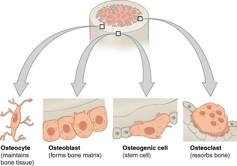
Bone contains a relatively small number of cells entrenched in a matrix of collagen fibers that provide a surface for inorganic salt crystals to adhere. These salt crystals form when calcium phosphate and calcium carbonate combine to create hydroxyapatite, which incorporates other inorganic salts like magnesium hydroxide, fluoride, and sulfate as it crystallizes, or calcifies, on the collagen fibers. The hydroxyapatite crystals give bones their hardness and strength, while the collagen fibers give them flexibility so that they are not brittle.

Although bone cells compose a small amount of the bone volume, they are crucial to the function of bones. Four types of cells are found within bone tissue: osteoblasts, osteocytes, osteogenic cells, and osteoclasts ( [link] ).

Bone cells

The top of this diagram shows the cross section of a generic bone with three zoom in boxes. The first box is on the periosteum. The second box is on the middle of the compact bone layer. The third box is on the inner edge of the compact bone where it transitions into the spongy bone. The callout in the periosteum points to two images. In the first image, four osteoblast cells are sitting end to end on the periosteum. The osteoblasts are roughly square shaped, except for one of the cells which is developing small, finger like projections. The caption says, “Osteoblasts form the matrix of the bone.” The second image called out from the periosteum shows a large, amorphous osteogenic cell sitting on the periosteum. The osteogenic cell is surrounded on both sides by a row of much smaller osteoblasts. The cell is shaped like a mushroom cap and also has finger like projections. The cell is a stem cell that develops into other bone cells. The box in the middle of the compact bone layer is pointing to an osteocyte. The osteocyte is a thin cell, roughly diamond shaped, with many branching, finger-like projections. The osteoctyes maintain bone tissue. The box at the inner edge of the compact bone is pointing to an osteoclast. The osteoclast is a large, round cell with multiple nuclei. It also has rows of fine finger like projections on its lower surface where it is sitting on the compact bone. The osteoclast reabsorbs bone.
Four types of cells are found within bone tissue. Osteogenic cells are undifferentiated and develop into osteoblasts. When osteoblasts get trapped within the calcified matrix, their structure and function changes, and they become osteocytes. Osteoclasts develop from monocytes and macrophages and differ in appearance from other bone cells.

Questions & Answers

what does preconceived mean
sammie Reply
physiological Psychology
Nwosu Reply
How can I develope my cognitive domain
Amanyire Reply
why is communication effective
Dakolo Reply
Communication is effective because it allows individuals to share ideas, thoughts, and information with others.
effective communication can lead to improved outcomes in various settings, including personal relationships, business environments, and educational settings. By communicating effectively, individuals can negotiate effectively, solve problems collaboratively, and work towards common goals.
it starts up serve and return practice/assessments.it helps find voice talking therapy also assessments through relaxed conversation.
miss
Every time someone flushes a toilet in the apartment building, the person begins to jumb back automatically after hearing the flush, before the water temperature changes. Identify the types of learning, if it is classical conditioning identify the NS, UCS, CS and CR. If it is operant conditioning, identify the type of consequence positive reinforcement, negative reinforcement or punishment
Wekolamo Reply
please i need answer
Wekolamo
because it helps many people around the world to understand how to interact with other people and understand them well, for example at work (job).
Manix Reply
Agreed 👍 There are many parts of our brains and behaviors, we really need to get to know. Blessings for everyone and happy Sunday!
ARC
A child is a member of community not society elucidate ?
JESSY Reply
Isn't practices worldwide, be it psychology, be it science. isn't much just a false belief of control over something the mind cannot truly comprehend?
Simon Reply
compare and contrast skinner's perspective on personality development on freud
namakula Reply
Skinner skipped the whole unconscious phenomenon and rather emphasized on classical conditioning
war
explain how nature and nurture affect the development and later the productivity of an individual.
Amesalu Reply
nature is an hereditary factor while nurture is an environmental factor which constitute an individual personality. so if an individual's parent has a deviant behavior and was also brought up in an deviant environment, observation of the behavior and the inborn trait we make the individual deviant.
Samuel
I am taking this course because I am hoping that I could somehow learn more about my chosen field of interest and due to the fact that being a PsyD really ignites my passion as an individual the more I hope to learn about developing and literally explore the complexity of my critical thinking skills
Zyryn Reply
good👍
Jonathan
and having a good philosophy of the world is like a sandwich and a peanut butter 👍
Jonathan
generally amnesi how long yrs memory loss
Kelu Reply
interpersonal relationships
Abdulfatai Reply
What would be the best educational aid(s) for gifted kids/savants?
Heidi Reply
treat them normal, if they want help then give them. that will make everyone happy
Saurabh
What are the treatment for autism?
Magret Reply
hello. autism is a umbrella term. autistic kids have different disorder overlapping. for example. a kid may show symptoms of ADHD and also learning disabilities. before treatment please make sure the kid doesn't have physical disabilities like hearing..vision..speech problem. sometimes these
Jharna
continue.. sometimes due to these physical problems..the diagnosis may be misdiagnosed. treatment for autism. well it depends on the severity. since autistic kids have problems in communicating and adopting to the environment.. it's best to expose the child in situations where the child
Jharna
child interact with other kids under doc supervision. play therapy. speech therapy. Engaging in different activities that activate most parts of the brain.. like drawing..painting. matching color board game. string and beads game. the more you interact with the child the more effective
Jharna
results you'll get.. please consult a therapist to know what suits best on your child. and last as a parent. I know sometimes it's overwhelming to guide a special kid. but trust the process and be strong and patient as a parent.
Jharna
Got questions? Join the online conversation and get instant answers!
Jobilize.com Reply

Get Jobilize Job Search Mobile App in your pocket Now!

Get it on Google Play Download on the App Store Now


Source:  OpenStax, Anatomy & Physiology. OpenStax CNX. Feb 04, 2016 Download for free at http://legacy.cnx.org/content/col11496/1.8
Google Play and the Google Play logo are trademarks of Google Inc.

Notification Switch

Would you like to follow the 'Anatomy & Physiology' conversation and receive update notifications?

Ask