<< Chapter < Page Chapter >> Page >
By the end of this section, you will be able to:
  • Identify the classes of cells that make up nervous tissue
  • Discuss how nervous tissue mediates perception and response

Nervous tissue is characterized as being excitable and capable of sending and receiving electrochemical signals that provide the body with information. Two main classes of cells make up nervous tissue: the neuron    and neuroglia    ( [link] ). Neurons propagate information via electrochemical impulses, called action potentials, which are biochemically linked to the release of chemical signals. Neuroglia play an essential role in supporting neurons and modulating their information propagation.

The neuron

This figure shows a diagram of a neuron and a micrograph showing two neuron cells. The body of the neuron contains a single, purple nucleus. The cell is irregularly shaped, having many projections emerging from its surface. Six sets of dendrites project from the top, right, and bottom edges of the cell. The dendrites are yellow and branch many times after leaving the cell, taking on the appearance of tiny trees. The axon projects from the left edge of the cell. The axon is a long cable like structure that branches into several finger like projections at its end. This is where the neuron makes contact with other cells. A label also notes that the area where the axon emerges from the cell body contains microfibrils and microtubules. The micrograph is considerably less magnified than the diagram. The neurons stain darkly and their nuclei are clearly visible. Their irregular cell body is also visible, along with the beginning of the axons.
The cell body of a neuron, also called the soma, contains the nucleus and mitochondria. The dendrites transfer the nerve impulse to the soma. The axon carries the action potential away to another excitable cell. LM × 1600. (Micrograph provided by the Regents of University of Michigan Medical School © 2012)

Follow this link to learn more about nervous tissue. What are the main parts of a nerve cell?

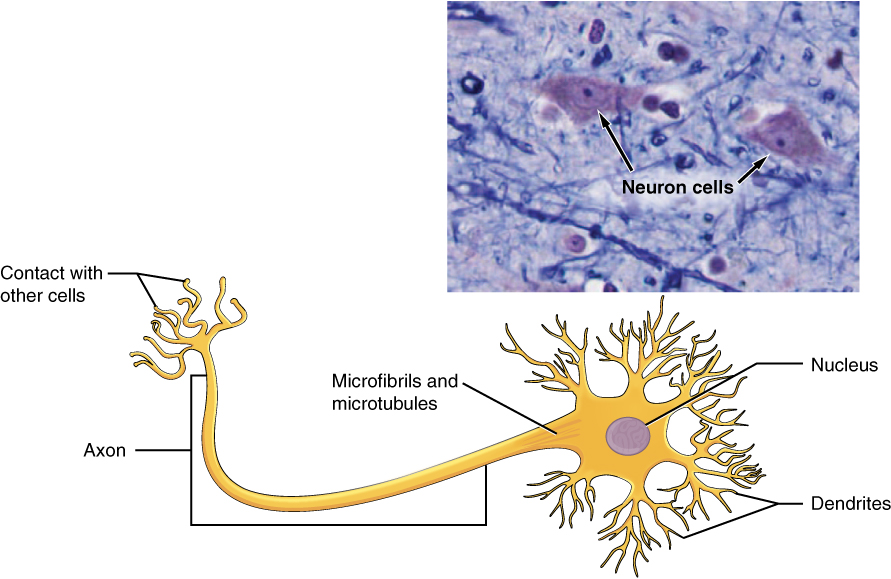
Neurons display distinctive morphology, well suited to their role as conducting cells, with three main parts. The cell body includes most of the cytoplasm, the organelles, and the nucleus. Dendrites branch off the cell body and appear as thin extensions. A long “tail,” the axon, extends from the neuron body and can be wrapped in an insulating layer known as myelin    , which is formed by accessory cells. The synapse is the gap between nerve cells, or between a nerve cell and its target, for example, a muscle or a gland, across which the impulse is transmitted by chemical compounds known as neurotransmitters. Neurons categorized as multipolar neurons have several dendrites and a single prominent axon. Bipolar neurons possess a single dendrite and axon with the cell body, while unipolar neurons have only a single process extending out from the cell body, which divides into a functional dendrite and into a functional axon. When a neuron is sufficiently stimulated, it generates an action potential that propagates down the axon towards the synapse. If enough neurotransmitters are released at the synapse to stimulate the next neuron or target, a response is generated.

The second class of neural cells comprises the neuroglia or glial cells, which have been characterized as having a simple support role. The word “glia” comes from the Greek word for glue. Recent research is shedding light on the more complex role of neuroglia in the function of the brain and nervous system. Astrocyte cells, named for their distinctive star shape, are abundant in the central nervous system. The astrocytes have many functions, including regulation of ion concentration in the intercellular space, uptake and/or breakdown of some neurotransmitters, and formation of the blood-brain barrier, the membrane that separates the circulatory system from the brain. Microglia protect the nervous system against infection but are not nervous tissue because they are related to macrophages. Oligodendrocyte cells produce myelin in the central nervous system (brain and spinal cord) while the Schwann cell    produces myelin in the peripheral nervous system ( [link] ).

Nervous tissue

Part A of this diagram shows various types of nerve cells. The largest cell is a neuron. The central body of the neuron contains a single nucleus. Six sets of dendrites project from the top, left and right, edges of the neuron. The dendrites are yellow and branch many times after leaving the cell, taking on the appearance of tiny trees. The axon projects from the bottom edge of the cell and is covered with purple sheaths labeled the myelin sheath. The sheath is not continuous, but instead is a series of equally spaced segments along the axon. Another cell, called an oligodendrocyte, is spider like in appearance, with its leg-like projections each connecting to a segment of the neuron’s myelin sheath. Above the neuron are three astrocytes. They are much smaller than the neuron and have no axons, and are also irregularly shaped cells with many dendrites projecting from the central body. Finally, a microglial cell is shown above the neuron. It is the smallest of the cells in this figure and is an elongated cell with many fine, tentacle-like projections. The projections are concentrated at the two ends of the cell, with the middle area lacking any projections. The micrograph of the neural tissue shows that this tissue is very heterogenous, with both large and small cells embedded in the matrix. Much of the space between the cells is occupied by threadlike nerve fibers.
Nervous tissue is made up of neurons and neuroglia. The cells of nervous tissue are specialized to transmit and receive impulses. LM × 872. (Micrograph provided by the Regents of University of Michigan Medical School © 2012)

Chapter review

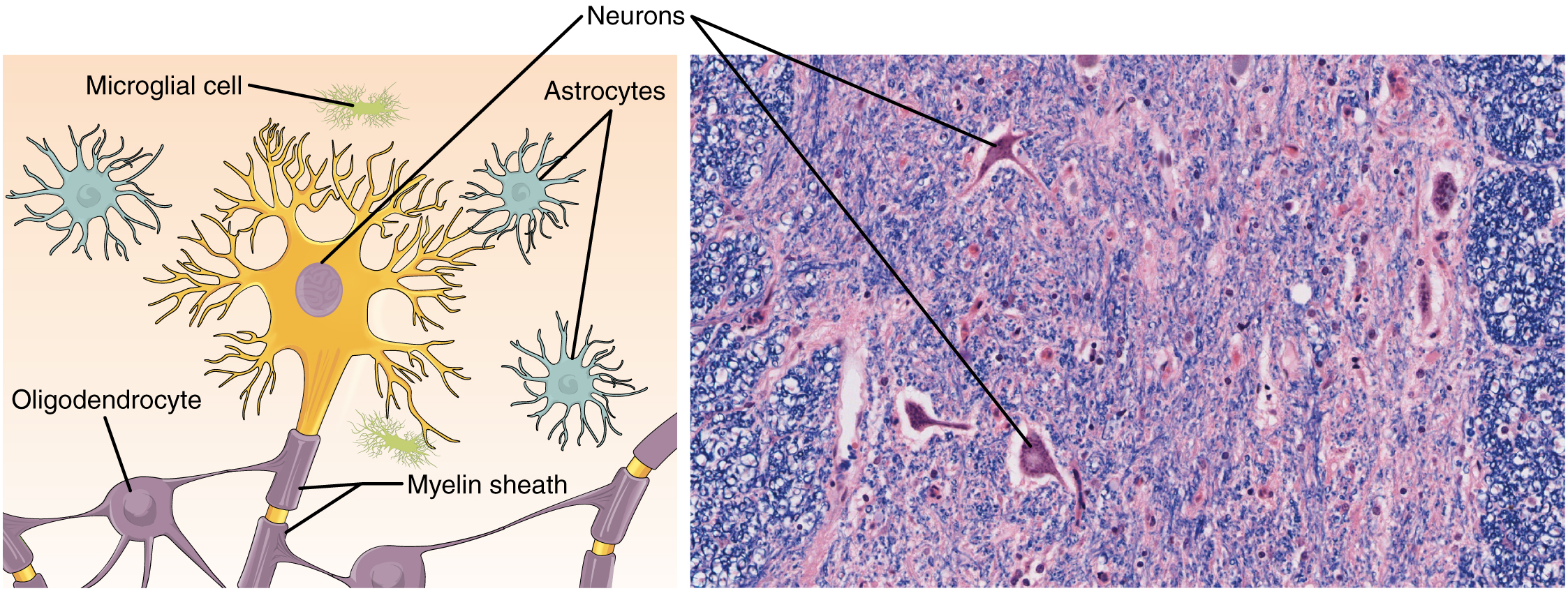
The most prominent cell of the nervous tissue, the neuron, is characterized mainly by its ability to receive stimuli and respond by generating an electrical signal, known as an action potential, which can travel rapidly over great distances in the body. A typical neuron displays a distinctive morphology: a large cell body branches out into short extensions called dendrites, which receive chemical signals from other neurons, and a long tail called an axon, which relays signals away from the cell to other neurons, muscles, or glands. Many axons are wrapped by a myelin sheath, a lipid derivative that acts as an insulator and speeds up the transmission of the action potential. Other cells in the nervous tissue, the neuroglia, include the astrocytes, microglia, oligodendrocytes, and Schwann cells.

Follow this link to learn more about nervous tissue. What are the main parts of a nerve cell?

Dendrites, cell body, and the axon.

Got questions? Get instant answers now!

References

Stern, P. Focus issue: getting excited about glia. Science [Internet]. 2010 [cited 2012 Dec 4]; 3(147):330-773. Available from:

(External Link)

Ming GL, Song H. Adult neurogenesis in the mammalian central nervous system. Annu. Rev. Neurosci. 2005 [cited 2012 Dec 4]; 28:223–250.

Questions & Answers

how does Neisseria cause meningitis
Nyibol Reply
what is microbiologist
Muhammad Reply
what is errata
Muhammad
is the branch of biology that deals with the study of microorganisms.
Ntefuni Reply
What is microbiology
Mercy Reply
studies of microbes
Louisiaste
when we takee the specimen which lumbar,spin,
Ziyad Reply
How bacteria create energy to survive?
Muhamad Reply
Bacteria doesn't produce energy they are dependent upon their substrate in case of lack of nutrients they are able to make spores which helps them to sustain in harsh environments
_Adnan
But not all bacteria make spores, l mean Eukaryotic cells have Mitochondria which acts as powerhouse for them, since bacteria don't have it, what is the substitution for it?
Muhamad
they make spores
Louisiaste
what is sporadic nd endemic, epidemic
Aminu Reply
the significance of food webs for disease transmission
Abreham
food webs brings about an infection as an individual depends on number of diseased foods or carriers dully.
Mark
explain assimilatory nitrate reduction
Esinniobiwa Reply
Assimilatory nitrate reduction is a process that occurs in some microorganisms, such as bacteria and archaea, in which nitrate (NO3-) is reduced to nitrite (NO2-), and then further reduced to ammonia (NH3).
Elkana
This process is called assimilatory nitrate reduction because the nitrogen that is produced is incorporated in the cells of microorganisms where it can be used in the synthesis of amino acids and other nitrogen products
Elkana
Examples of thermophilic organisms
Shu Reply
Give Examples of thermophilic organisms
Shu
advantages of normal Flora to the host
Micheal Reply
Prevent foreign microbes to the host
Abubakar
they provide healthier benefits to their hosts
ayesha
They are friends to host only when Host immune system is strong and become enemies when the host immune system is weakened . very bad relationship!
Mark
what is cell
faisal Reply
cell is the smallest unit of life
Fauziya
cell is the smallest unit of life
Akanni
ok
Innocent
cell is the structural and functional unit of life
Hasan
is the fundamental units of Life
Musa
what are emergency diseases
Micheal Reply
There are nothing like emergency disease but there are some common medical emergency which can occur simultaneously like Bleeding,heart attack,Breathing difficulties,severe pain heart stock.Hope you will get my point .Have a nice day ❣️
_Adnan
define infection ,prevention and control
Innocent
I think infection prevention and control is the avoidance of all things we do that gives out break of infections and promotion of health practices that promote life
Lubega
Heyy Lubega hussein where are u from?
_Adnan
en français
Adama
which site have a normal flora
ESTHER Reply
Many sites of the body have it Skin Nasal cavity Oral cavity Gastro intestinal tract
Safaa
skin
Asiina
skin,Oral,Nasal,GIt
Sadik
How can Commensal can Bacteria change into pathogen?
Sadik
How can Commensal Bacteria change into pathogen?
Sadik
all
Tesfaye
by fussion
Asiina
what are the advantages of normal Flora to the host
Micheal
what are the ways of control and prevention of nosocomial infection in the hospital
Micheal
what is inflammation
Shelly Reply
part of a tissue or an organ being wounded or bruised.
Wilfred
what term is used to name and classify microorganisms?
Micheal Reply
Binomial nomenclature
adeolu
Got questions? Join the online conversation and get instant answers!
Jobilize.com Reply

Get Jobilize Job Search Mobile App in your pocket Now!

Get it on Google Play Download on the App Store Now


Source:  OpenStax, Anatomy & Physiology. OpenStax CNX. Feb 04, 2016 Download for free at http://legacy.cnx.org/content/col11496/1.8
Google Play and the Google Play logo are trademarks of Google Inc.

Notification Switch

Would you like to follow the 'Anatomy & Physiology' conversation and receive update notifications?

Ask