

| << Chapter < Page | Chapter >> Page > |
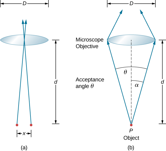
where d is the distance between the specimen and the objective lens, and we have used the small angle approximation (i.e., we have assumed that x is much smaller than d ), so that Therefore, the resolving power is
Another way to look at this is by the concept of numerical aperture ( NA ), which is a measure of the maximum acceptance angle at which a lens will take light and still contain it within the lens. [link] (b) shows a lens and an object at point P . The NA here is a measure of the ability of the lens to gather light and resolve fine detail. The angle subtended by the lens at its focus is defined to be . From the figure and again using the small angle approximation, we can write
The NA for a lens is , where n is the index of refraction of the medium between the objective lens and the object at point P . From this definition for NA , we can see that
In a microscope, NA is important because it relates to the resolving power of a lens. A lens with a large NA is able to resolve finer details. Lenses with larger NA are also able to collect more light and so give a brighter image. Another way to describe this situation is that the larger the NA , the larger the cone of light that can be brought into the lens, so more of the diffraction modes are collected. Thus the microscope has more information to form a clear image, and its resolving power is higher.
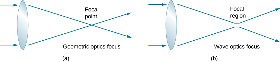
One of the consequences of diffraction is that the focal point of a beam has a finite width and intensity distribution. Imagine focusing when only considering geometric optics, as in [link] (a). The focal point is regarded as an infinitely small point with a huge intensity and the capacity to incinerate most samples, irrespective of the NA of the objective lens—an unphysical oversimplification. For wave optics, due to diffraction, we take into account the phenomenon in which the focal point spreads to become a focal spot ( [link] (b)) with the size of the spot decreasing with increasing NA . Consequently, the intensity in the focal spot increases with increasing NA . The higher the NA , the greater the chances of photodegrading the specimen. However, the spot never becomes a true point.
In a different type of microscope, molecules within a specimen are made to emit light through a mechanism called fluorescence. By controlling the molecules emitting light, it has become possible to construct images with resolution much finer than the Rayleigh criterion, thus circumventing the diffraction limit. The development of super-resolved fluorescence microscopy led to the 2014 Nobel Prize in Chemistry.
Notification Switch
Would you like to follow the 'University physics volume 3' conversation and receive update notifications?

|
|

|
|

|
|

|
|

|
|

|
|

|
|

|
|

|
|

|