<< Chapter < Page Chapter >> Page >
By the end of this section, you will be able to:
  • Describe the combined effect of interference and diffraction with two slits, each with finite width
  • Determine the relative intensities of interference fringes within a diffraction pattern
  • Identify missing orders, if any

When we studied interference in Young’s double-slit experiment, we ignored the diffraction effect in each slit. We assumed that the slits were so narrow that on the screen you saw only the interference of light from just two point sources. If the slit is smaller than the wavelength, then [link] (a) shows that there is just a spreading of light and no peaks or troughs on the screen. Therefore, it was reasonable to leave out the diffraction effect in that chapter. However, if you make the slit wider, [link] (b) and (c) show that you cannot ignore diffraction. In this section, we study the complications to the double-slit experiment that arise when you also need to take into account the diffraction effect of each slit.

To calculate the diffraction pattern for two (or any number of) slits, we need to generalize the method we just used for a single slit. That is, across each slit, we place a uniform distribution of point sources that radiate Huygens wavelets, and then we sum the wavelets from all the slits. This gives the intensity at any point on the screen. Although the details of that calculation can be complicated, the final result is quite simple:

Two-slit diffraction pattern

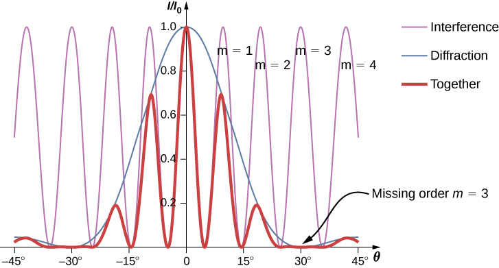
The diffraction pattern of two slits of width D that are separated by a distance d is the interference pattern of two point sources separated by d multiplied by the diffraction pattern of a slit of width D .

In other words, the locations of the interference fringes are given by the equation d sin θ = m λ , the same as when we considered the slits to be point sources, but the intensities of the fringes are now reduced by diffraction effects, according to [link] . [Note that in the chapter on interference, we wrote d sin θ = m λ and used the integer m to refer to interference fringes. [link] also uses m , but this time to refer to diffraction minima. If both equations are used simultaneously, it is good practice to use a different variable (such as n ) for one of these integers in order to keep them distinct.]

Interference and diffraction effects operate simultaneously and generally produce minima at different angles. This gives rise to a complicated pattern on the screen, in which some of the maxima of interference from the two slits are missing if the maximum of the interference is in the same direction as the minimum of the diffraction. We refer to such a missing peak as a missing order    . One example of a diffraction pattern on the screen is shown in [link] . The solid line with multiple peaks of various heights is the intensity observed on the screen. It is a product of the interference pattern of waves from separate slits and the diffraction of waves from within one slit.

Figure shows a graph of I by I0 versus theta. Three curves are shown on the graph. Interference curve has a smaller wavelength. Diffraction curve has a larger wavelength and a y value of 1 at x equal to 0. Resultant curve has the same wavelength as the interference and its amplitude is modified according to the diffraction curve. Each wave crest of the interference wave is labeled m equal to 1, m equal to 2 and so on. The diffraction wave has a zero at m equal to 3, and theta equal to 30 degrees. Hence, the resultant wave, too, has a zero. This is labeled missing order m equal to 3.
Diffraction from a double slit. The purple line with peaks of the same height are from the interference of the waves from two slits; the blue line with one big hump in the middle is the diffraction of waves from within one slit; and the thick red line is the product of the two, which is the pattern observed on the screen. The plot shows the expected result for a slit width D = 2 λ and slit separation d = 6 λ . The maximum of m = ± 3 order for the interference is missing because the minimum of the diffraction occurs in the same direction.

Questions & Answers

what is phylogeny
Odigie Reply
evolutionary history and relationship of an organism or group of organisms
AI-Robot
ok
Deng
what is biology
Hajah Reply
the study of living organisms and their interactions with one another and their environments
AI-Robot
what is biology
Victoria Reply
HOW CAN MAN ORGAN FUNCTION
Alfred Reply
the diagram of the digestive system
Assiatu Reply
allimentary cannel
Ogenrwot
How does twins formed
William Reply
They formed in two ways first when one sperm and one egg are splited by mitosis or two sperm and two eggs join together
Oluwatobi
what is genetics
Josephine Reply
Genetics is the study of heredity
Misack
how does twins formed?
Misack
What is manual
Hassan Reply
discuss biological phenomenon and provide pieces of evidence to show that it was responsible for the formation of eukaryotic organelles
Joseph Reply
what is biology
Yousuf Reply
the study of living organisms and their interactions with one another and their environment.
Wine
discuss the biological phenomenon and provide pieces of evidence to show that it was responsible for the formation of eukaryotic organelles in an essay form
Joseph Reply
what is the blood cells
Shaker Reply
list any five characteristics of the blood cells
Shaker
lack electricity and its more savely than electronic microscope because its naturally by using of light
Abdullahi Reply
advantage of electronic microscope is easily and clearly while disadvantage is dangerous because its electronic. advantage of light microscope is savely and naturally by sun while disadvantage is not easily,means its not sharp and not clear
Abdullahi
cell theory state that every organisms composed of one or more cell,cell is the basic unit of life
Abdullahi
is like gone fail us
DENG
cells is the basic structure and functions of all living things
Ramadan
What is classification
ISCONT Reply
is organisms that are similar into groups called tara
Yamosa
in what situation (s) would be the use of a scanning electron microscope be ideal and why?
Kenna Reply
A scanning electron microscope (SEM) is ideal for situations requiring high-resolution imaging of surfaces. It is commonly used in materials science, biology, and geology to examine the topography and composition of samples at a nanoscale level. SEM is particularly useful for studying fine details,
Hilary
Got questions? Join the online conversation and get instant answers!
Jobilize.com Reply
Practice Key Terms 2

Get Jobilize Job Search Mobile App in your pocket Now!

Get it on Google Play Download on the App Store Now


Source:  OpenStax, University physics volume 3. OpenStax CNX. Nov 04, 2016 Download for free at http://cnx.org/content/col12067/1.4
Google Play and the Google Play logo are trademarks of Google Inc.

Notification Switch

Would you like to follow the 'University physics volume 3' conversation and receive update notifications?

Ask