<< Chapter < Page Chapter >> Page >
By the end of this section, you will be able to:
  • Explain what a continuous source charge distribution is and how it is related to the concept of quantization of charge
  • Describe line charges, surface charges, and volume charges
  • Calculate the field of a continuous source charge distribution of either sign

The charge distributions we have seen so far have been discrete: made up of individual point particles. This is in contrast with a continuous charge distribution    , which has at least one nonzero dimension. If a charge distribution is continuous rather than discrete, we can generalize the definition of the electric field. We simply divide the charge into infinitesimal pieces and treat each piece as a point charge.

Note that because charge is quantized, there is no such thing as a “truly” continuous charge distribution. However, in most practical cases, the total charge creating the field involves such a huge number of discrete charges that we can safely ignore the discrete nature of the charge and consider it to be continuous. This is exactly the kind of approximation we make when we deal with a bucket of water as a continuous fluid, rather than a collection of H 2 O molecules.

Our first step is to define a charge density for a charge distribution along a line, across a surface, or within a volume, as shown in [link] .

Figure a shows a long rod with linear charge density lambda. A small segment of the rod is shaded and labeled d l. Figure b shows a surface with surface charge density sigma. A small area within the surface is shaded and labeled d A. Figure c shows a volume with volume charge density rho. A small volume within it is shaded and labeled d V. Figure d shows a surface with two regions shaded and labeled q 1 and q2. A point P is identified above (not on) the surface. A thin line indicates the distance from each of the shaded regions. The vectors E 1 and E 2 are drawn at point P and point away from the respective shaded region. E net is the vector sum of E 1 and E 2. In this case, it points up, away from the surface.
The configuration of charge differential elements for a (a) line charge, (b) sheet of charge, and (c) a volume of charge. Also note that (d) some of the components of the total electric field cancel out, with the remainder resulting in a net electric field.

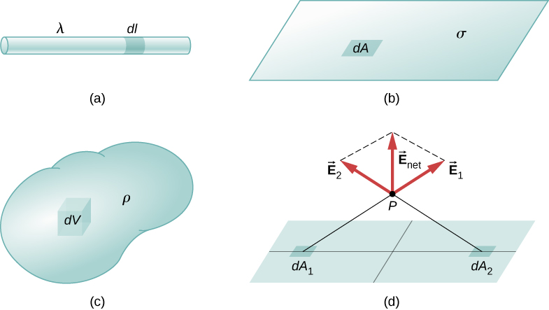
Definitions of charge density:

  • λ charge per unit length ( linear charge density    ); units are coulombs per meter (C/m)
  • σ charge per unit area ( surface charge density    ); units are coulombs per square meter ( C / m 2 )
  • ρ charge per unit volume ( volume charge density    ); units are coulombs per cubic meter ( C / m 3 )

Then, for a line charge, a surface charge, and a volume charge, the summation in [link] becomes an integral and q i is replaced by d q = λ d l , σ d A , or ρ d V , respectively:

Point charge: E ( P ) = 1 4 π ε 0 i = 1 N ( q i r 2 ) r ^
Line charge: E ( P ) = 1 4 π ε 0 line ( λ d l r 2 ) r ^
Surface charge: E ( P ) = 1 4 π ε 0 surface ( σ d A r 2 ) r ^
Volume charge: E ( P ) = 1 4 π ε 0 volume ( ρ d V r 2 ) r ^

The integrals are generalizations of the expression for the field of a point charge. They implicitly include and assume the principle of superposition. The “trick” to using them is almost always in coming up with correct expressions for dl , dA , or dV , as the case may be, expressed in terms of r , and also expressing the charge density function appropriately. It may be constant; it might be dependent on location.

Note carefully the meaning of r in these equations: It is the distance from the charge element ( q i , λ d l , σ d A , ρ d V ) to the location of interest, P ( x , y , z ) (the point in space where you want to determine the field). However, don’t confuse this with the meaning of r ^ ; we are using it and the vector notation E to write three integrals at once. That is, [link] is actually

E x ( P ) = 1 4 π ε 0 line ( λ d l r 2 ) x , E y ( P ) = 1 4 π ε 0 line ( λ d l r 2 ) y , E z ( P ) = 1 4 π ε 0 line ( λ d l r 2 ) z .

Questions & Answers

what is phylogeny
Odigie Reply
evolutionary history and relationship of an organism or group of organisms
AI-Robot
ok
Deng
what is biology
Hajah Reply
the study of living organisms and their interactions with one another and their environments
AI-Robot
what is biology
Victoria Reply
HOW CAN MAN ORGAN FUNCTION
Alfred Reply
the diagram of the digestive system
Assiatu Reply
allimentary cannel
Ogenrwot
How does twins formed
William Reply
They formed in two ways first when one sperm and one egg are splited by mitosis or two sperm and two eggs join together
Oluwatobi
what is genetics
Josephine Reply
Genetics is the study of heredity
Misack
how does twins formed?
Misack
What is manual
Hassan Reply
discuss biological phenomenon and provide pieces of evidence to show that it was responsible for the formation of eukaryotic organelles
Joseph Reply
what is biology
Yousuf Reply
the study of living organisms and their interactions with one another and their environment.
Wine
discuss the biological phenomenon and provide pieces of evidence to show that it was responsible for the formation of eukaryotic organelles in an essay form
Joseph Reply
what is the blood cells
Shaker Reply
list any five characteristics of the blood cells
Shaker
lack electricity and its more savely than electronic microscope because its naturally by using of light
Abdullahi Reply
advantage of electronic microscope is easily and clearly while disadvantage is dangerous because its electronic. advantage of light microscope is savely and naturally by sun while disadvantage is not easily,means its not sharp and not clear
Abdullahi
cell theory state that every organisms composed of one or more cell,cell is the basic unit of life
Abdullahi
is like gone fail us
DENG
cells is the basic structure and functions of all living things
Ramadan
What is classification
ISCONT Reply
is organisms that are similar into groups called tara
Yamosa
in what situation (s) would be the use of a scanning electron microscope be ideal and why?
Kenna Reply
A scanning electron microscope (SEM) is ideal for situations requiring high-resolution imaging of surfaces. It is commonly used in materials science, biology, and geology to examine the topography and composition of samples at a nanoscale level. SEM is particularly useful for studying fine details,
Hilary
Got questions? Join the online conversation and get instant answers!
Jobilize.com Reply
Practice Key Terms 6

Get Jobilize Job Search Mobile App in your pocket Now!

Get it on Google Play Download on the App Store Now


Source:  OpenStax, University physics volume 2. OpenStax CNX. Oct 06, 2016 Download for free at http://cnx.org/content/col12074/1.3
Google Play and the Google Play logo are trademarks of Google Inc.

Notification Switch

Would you like to follow the 'University physics volume 2' conversation and receive update notifications?

Ask