

| << Chapter < Page | Chapter >> Page > |
We have seen that under certain circumstances particles behave like waves. This idea is used in the electron microscope which is a type of microscope that uses electrons to create an image of the target. It has much higher magnification or resolving power than a normal light microscope, up to two million times, allowing it to see smaller objects and details.
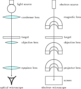
Let's first review how a regular optical microscope works. A beam of light is shone through a thin target and the image is then magnified and focused using objective and ocular lenses. The amount of light which passes through the target depends on the densities of the target since the less dense regions allow more light to pass through than the denser regions. This means that the beam of light which is partially transmitted through the target carries information about the inner structure of the target.
The original form of the electron microscopy, transmission electron microscopy, works in a similar manner using electrons. In the electron microscope, electrons which are emitted by a cathode are formed into a beam using magnetic lenses. This electron beam is then passed through a very thin target. Again, the regions in the target with higher densities stop the electrons more easily. So, the number of electrons which pass through the different regions of the target depends on their densities. This means that the partially transmitted beam of electrons carries information about the densities of the inner structure of the target. The spatial variation in this information (the "image") is then magnified by a series of magnetic lenses and it is recorded by hitting a fluorescent screen, photographic plate, or light sensitive sensor such as a CCD (charge-coupled device) camera. The image detected by the CCD may be displayed in real time on a monitor or computer. In [link] is an image of the polio virus obtained with a transmission electron microscope.

The structure of an optical and electron microscope are compared in [link] . While the optical microscope uses light and focuses using lenses, the electron microscope uses electrons and focuses using electromagnets.
Electron microscopes are very useful as they are able to magnify objects to a much higher resolution. This is because their de Broglie wavelengths are so much smaller than that of visible light. You hopefully remember that light is diffracted by objects which are separated by a distance of about the same size as the wavelength of the light. This diffraction then prevents you from being able to focus the transmitted light into an image. So the sizes at which diffraction occurs for a beam of electrons is much smaller than those for visible light. This is why you can magnify targets to a much higher order of magnification using electrons rather than visible light.
| Light microscope | Electron microscope | |
| Source | Bright lamp or laser | Electron gun |
| Radiation | U.V. or visible light | Electron beam produced by heating metal surface (e.g. tungsten) |
| Lenses | Curved glass surfaces | Electromagnets |
| Receiver | Eye; photographic emulsion or digital image | Fluorescent screen (for location and focusing image); photographic emulsion or digital image |
| Focus | Axial movement of lenses (up and down) | Adjustment of magnetic field in the electromagnets by changing the current |
| Operating | Atmospheric | High vacuum |
| Pressure |
Notification Switch
Would you like to follow the 'Siyavula textbooks: grade 12 physical science' conversation and receive update notifications?

|
|

|
|

|
|

|
|

|
|

|
|
|
|
|

|
|

|
|

|