

| << Chapter < Page | Chapter >> Page > |
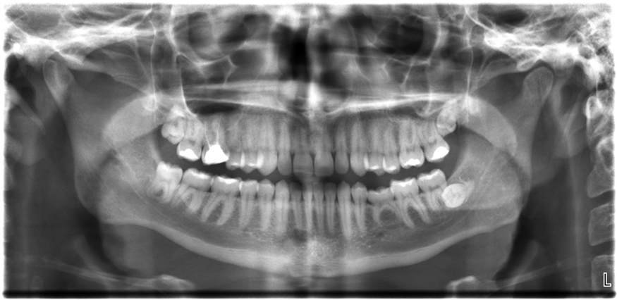
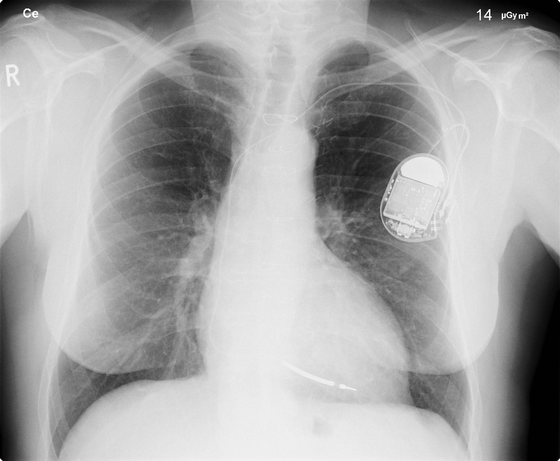
The most common x-ray images are simple shadows. Since x-ray photons have high energies, they penetrate materials that are opaque to visible light. The more energy an x-ray photon has, the more material it will penetrate. So an x-ray tube may be operated at 50.0 kV for a chest x ray, whereas it may need to be operated at 100 kV to examine a broken leg in a cast. The depth of penetration is related to the density of the material as well as to the energy of the photon. The denser the material, the fewer x-ray photons get through and the darker the shadow. Thus x rays excel at detecting breaks in bones and in imaging other physiological structures, such as some tumors, that differ in density from surrounding material. Because of their high photon energy, x rays produce significant ionization in materials and damage cells in biological organisms. Modern uses minimize exposure to the patient and eliminate exposure to others. Biological effects of x rays will be explored in the next chapter along with other types of ionizing radiation such as those produced by nuclei.
As the x-ray energy increases, the Compton effect (see Photon Momentum ) becomes more important in the attenuation of the x rays. Here, the x ray scatters from an outer electron shell of the atom, giving the ejected electron some kinetic energy while losing energy itself. The probability for attenuation of the x rays depends upon the number of electrons present (the material’s density) as well as the thickness of the material. Chemical composition of the medium, as characterized by its atomic number , is not important here. Low-energy x rays provide better contrast (sharper images). However, due to greater attenuation and less scattering, they are more absorbed by thicker materials. Greater contrast can be achieved by injecting a substance with a large atomic number, such as barium or iodine. The structure of the part of the body that contains the substance (e.g., the gastro-intestinal tract or the abdomen) can easily be seen this way.
Breast cancer is the second-leading cause of death among women worldwide. Early detection can be very effective, hence the importance of x-ray diagnostics. A mammogram cannot diagnose a malignant tumor, only give evidence of a lump or region of increased density within the breast. X-ray absorption by different types of soft tissue is very similar, so contrast is difficult; this is especially true for younger women, who typically have denser breasts. For older women who are at greater risk of developing breast cancer, the presence of more fat in the breast gives the lump or tumor more contrast. MRI (Magnetic resonance imaging) has recently been used as a supplement to conventional x rays to improve detection and eliminate false positives. The subject’s radiation dose from x rays will be treated in a later chapter.
Notification Switch
Would you like to follow the 'College physics' conversation and receive update notifications?

|
|
|
|
|

|
|

|
|

|
|

|
|
|
|
|

|
|
|
|
|

|
|

|
|

|