<< Chapter < Page Chapter >> Page >

Below the epidermis of eudicot leaves are layers of cells known as the mesophyll, or “middle leaf.” The mesophyll of most leaves typically contains two arrangements of parenchyma cells: the palisade parenchyma and spongy parenchyma ( [link] ). The palisade parenchyma (also called the palisade mesophyll) has column-shaped, tightly packed cells, and may be present in one, two, or three layers. Below the palisade parenchyma are loosely arranged cells of an irregular shape. These are the cells of the spongy parenchyma (or spongy mesophyll). The air space found between the spongy parenchyma cells allows gaseous exchange between the leaf and the outside atmosphere through the stomata. In aquatic plants, the intercellular spaces in the spongy parenchyma help the leaf float. Both layers of the mesophyll contain many chloroplasts. Guard cells are the only epidermal cells to contain chloroplasts.

 Part A is a leaf cross section illustration. A flat layer of rectangular cells make up the upper and lower epidermis. A cuticle layer protects the outside of both epidermal layers. A stomatal pore in the lower epidermis allows carbon dioxide to enter and oxygen to leave. Oval guard cells surround the pore. Sandwiched between the upper and lower epidermis is the mesophyll. The upper part of the mesophyll is comprised of columnar cells called palisade parenchyma. The lower part of the mesophyll is made up of loosely packed spongy parenchyma. Part B is a scanning electron micrograph of a leaf in which all the layers described above are visible. Palisade cells are about 50 microns tall and 10 microns wide and are covered with tiny bumps, which are the chloroplasts. Spongy cells smaller and irregularly shaped. Several large bumps about 20 microns across project from the lower surface of the leaf.
In the (a) leaf drawing, the central mesophyll is sandwiched between an upper and lower epidermis. The mesophyll has two layers: an upper palisade layer comprised of tightly packed, columnar cells, and a lower spongy layer, comprised of loosely packed, irregularly shaped cells. Stomata on the leaf underside allow gas exchange. A waxy cuticle covers all aerial surfaces of land plants to minimize water loss. These leaf layers are clearly visible in the (b) scanning electron micrograph. The numerous small bumps in the palisade parenchyma cells are chloroplasts. Chloroplasts are also present in the spongy parenchyma, but are not as obvious. The bumps protruding from the lower surface of the leave are glandular trichomes, which differ in structure from the stalked trichomes in [link] . (credit b: modification of work by Robert R. Wise)

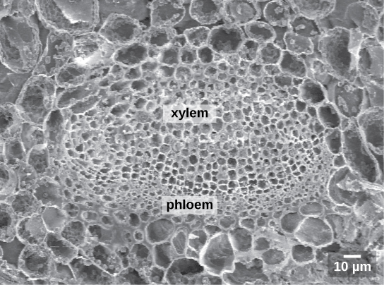
Like the stem, the leaf contains vascular bundles composed of xylem and phloem ( [link] ). The xylem consists of tracheids and vessels, which transport water and minerals to the leaves. The phloem transports the photosynthetic products from the leaf to the other parts of the plant. A single vascular bundle, no matter how large or small, always contains both xylem and phloem tissues.

 The scanning electron micrograph shows an oval vascular bundle. Small phloem cells make up the bottom of the bundle, and larger xylem cells make up the top. The bundle is surrounded by a ring of larger cells.
This scanning electron micrograph shows xylem and phloem in the leaf vascular bundle from the lyre-leaved sand cress ( Arabidopsis lyrata) . (credit: modification of work by Robert R. Wise; scale-bar data from Matt Russell)

Leaf adaptations

Coniferous plant species that thrive in cold environments, like spruce, fir, and pine, have leaves that are reduced in size and needle-like in appearance. These needle-like leaves have sunken stomata and a smaller surface area: two attributes that aid in reducing water loss. In hot climates, plants such as cacti have succulent leaves that help to conserve water. Many aquatic plants have leaves with wide lamina that can float on the surface of the water, and a thick waxy cuticle on the leaf surface that repels water.

Evolution connection

Plant adaptations in resource-deficient environments

Roots, stems, and leaves are structured to ensure that a plant can obtain the required resources of sunlight, water, soil nutrients, carbon dioxide and oxygen. Some remarkable adaptations have evolved to enable plant species to thrive in less than ideal habitats, where one or more of these resources is in short supply.

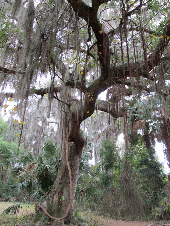
In tropical rainforests, light is often scarce, since many trees and plants grow close together and block much of the sunlight from reaching the forest floor. Many tropical plant species have exceptionally broad leaves to maximize the capture of sunlight. Other species are epiphytes: plants that grow on other plants that serve as a physical support. Such plants are able to grow high up in the canopy atop the branches of other trees, where sunlight is more plentiful. Epiphytes live on rain and minerals collected in the branches and leaves of the supporting plant. Bromeliads (members of the pineapple family), ferns, and orchids are examples of tropical epiphytes ( [link] ). Many epiphytes have specialized tissues that enable them to efficiently capture and store water.

Photo shows long, thin brown leaves of Spanish moss hanging down from the branches of a large oak tree.
One of the most well known bromeliads is Spanish moss ( Tillandsia usneoides ), seen here in an oak tree. (credit: Kristine Paulus)

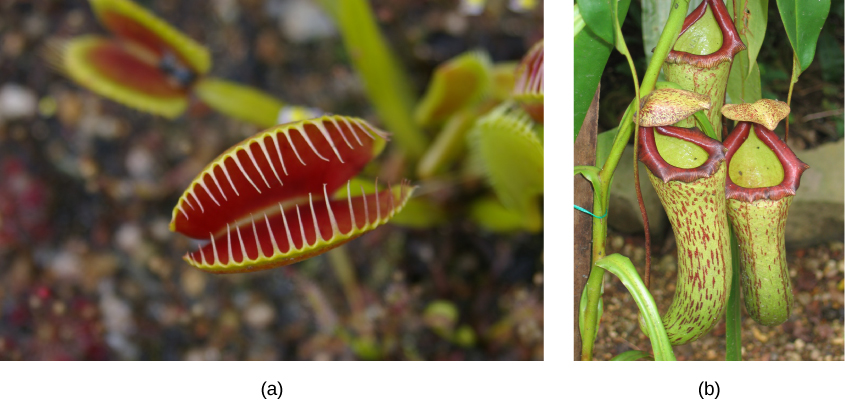
Some plants have special adaptations that help them to survive in nutrient-poor environments. Carnivorous plants, such as the Venus flytrap and the pitcher plant ( [link] ), grow in bogs where the soil is low in nitrogen. In these plants, leaves are modified to capture insects. The insect-capturing leaves may have evolved to provide these plants with a supplementary source of much-needed nitrogen.

 Left photo shows modified leaves of a Venus flytrap. The two leaves resemble the upper and lower part of the mouth, and are red on the interior. Hair-like appendages, like teeth, frame each modified leaf, so that when the leaves close, the insect will be trapped. Right photo shows three modified leaves of the pitcher plant, which are green tubes with red specks and have a red rim forming the top opening.
The (a) Venus flytrap has modified leaves that can capture insects. When an unlucky insect touches the trigger hairs inside the leaf, the trap suddenly closes. The opening of the (b) pitcher plant is lined with a slippery wax. Insects crawling on the lip slip and fall into a pool of water in the bottom of the pitcher, where they are digested by bacteria. The plant then absorbs the smaller molecules. (credit a: modification of work by Peter Shanks; credit b: modification of work by Tim Mansfield)

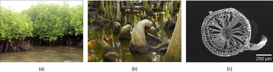
Many swamp plants have adaptations that enable them to thrive in wet areas, where their roots grow submerged underwater. In these aquatic areas, the soil is unstable and little oxygen is available to reach the roots. Trees such as mangroves ( Rhizophora sp.) growing in coastal waters produce aboveground roots that help support the tree ( [link] ). Some species of mangroves, as well as cypress trees, have pneumatophores: upward-growing roots containing pores and pockets of tissue specialized for gas exchange. Wild rice is an aquatic plant with large air spaces in the root cortex. The air-filled tissue—called aerenchyma—provides a path for oxygen to diffuse down to the root tips, which are embedded in oxygen-poor bottom sediments.

 Photo A shows mangrove trees with roots extending into the water. Part B shows cypress trees growing in the water, with upward-growing roots between the trees. Part C is a scanning electron micrograph showing a cross section of wild rice. The cells radiate from the center like spokes on a bicycle wheel, and are interspersed by large spaces that hold air.
The branches of (a) mangrove trees develop aerial roots, which descend to the ground and help to anchor the trees. (b) Cypress trees and some mangrove species have upward-growing roots called pneumatophores that are involved in gas exchange. Aquatic plants such as (c) wild rice have large spaces in the root cortex called aerenchyma, visualized here using scanning electron microscopy. (credit a: modification of work by Roberto Verzo; credit b: modification of work by Duane Burdick; credit c: modification of work by Robert R. Wise)

Get Jobilize Job Search Mobile App in your pocket Now!

Get it on Google Play Download on the App Store Now


Source:  OpenStax, Principles of biology. OpenStax CNX. Aug 09, 2016 Download for free at http://legacy.cnx.org/content/col11569/1.25
Google Play and the Google Play logo are trademarks of Google Inc.

Notification Switch

Would you like to follow the 'Principles of biology' conversation and receive update notifications?

Ask