

| << Chapter < Page | Chapter >> Page > |
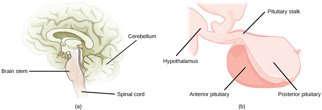
The pituitary gland , sometimes called the hypophysis or “master gland” is located at the base of the brain in the sella turcica, a groove of the sphenoid bone of the skull, illustrated in [link] . It is attached to the hypothalamus via a stalk called the pituitary stalk (or infundibulum). The anterior portion of the pituitary gland is regulated by releasing or release-inhibiting hormones produced by the hypothalamus, and the posterior pituitary receives signals via neurosecretory cells to release hormones produced by the hypothalamus. The pituitary has two distinct regions—the anterior pituitary and the posterior pituitary—which between them secrete nine different peptide or protein hormones. The posterior lobe of the pituitary gland contains axons of the hypothalamic neurons.
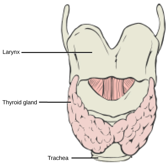
The thyroid gland is located in the neck, just below the larynx and in front of the trachea, as shown in [link] . It is a butterfly-shaped gland with two lobes that are connected by the isthmus. It has a dark red color due to its extensive vascular system. When the thyroid swells due to dysfunction, it can be felt under the skin of the neck.
Thyroid follicle cells synthesize the hormone thyroxine, which is also known as T 4 because it contains four atoms of iodine, and triiodothyronine, also known as T 3 because it contains three atoms of iodine. Follicle cells are stimulated to release stored T 3 and T 4 by thyroid stimulating hormone (TSH), which is produced by the anterior pituitary. These thyroid hormones increase the rates of mitochondrial ATP production.
A third hormone, calcitonin, is produced by parafollicular cells of the thyroid either releasing hormones or inhibiting hormones. Calcitonin release is not controlled by TSH, but instead is released when calcium ion concentrations in the blood rise. Calcitonin functions to help regulate calcium concentrations in body fluids. It acts in the bones to inhibit osteoclast activity and in the kidneys to stimulate excretion of calcium. The combination of these two events lowers body fluid levels of calcium.
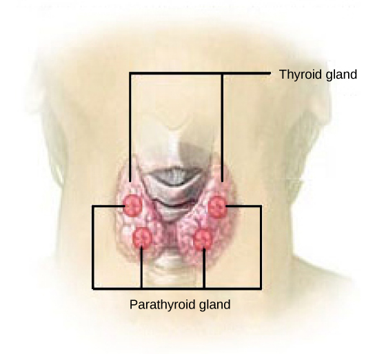
Most people have four parathyroid glands ; however, the number can vary from two to six. These glands are located on the posterior surface of the thyroid gland, as shown in [link] . Normally, there is a superior gland and an inferior gland associated with each of the thyroid’s two lobes. Each parathyroid gland is covered by connective tissue and contains many secretory cells that are associated with a capillary network.
The parathyroid glands produce parathyroid hormone (PTH). PTH increases blood calcium concentrations when calcium ion levels fall below normal. PTH (1) enhances reabsorption of Ca 2+ by the kidneys, (2) stimulates osteoclast activity and inhibits osteoblast activity, and (3) it stimulates synthesis and secretion of calcitriol by the kidneys, which enhances Ca 2+ absorption by the digestive system. PTH and calcitonin work in opposition to one another to maintain homeostatic Ca 2+ levels in body fluids.
Notification Switch
Would you like to follow the 'Principles of biology' conversation and receive update notifications?

|
|

|
|

|
|

|
|

|
|

|
|

|
|

|
|

|
|

|