

| << Chapter < Page | Chapter >> Page > |
Watch this video to learn more about the flexion and extension of the knee, as the femur both rolls and glides on the tibia to maintain stable contact between the bones in all knee positions. The patella glides along a groove on the anterior side of the distal femur. The collateral ligaments on the sides of the knee become tight in the fully extended position to help stabilize the knee. The posterior cruciate ligament supports the knee when flexed and the anterior cruciate ligament becomes tight when the knee comes into full extension to resist hyperextension. What are the ligaments that support the knee joint?
Watch this video to learn more about the anatomy of the knee joint, including bones, joints, muscles, nerves, and blood vessels. Which ligament of the knee keeps the tibia from sliding too far forward in relation to the femur and which ligament keeps the tibia from sliding too far backward?
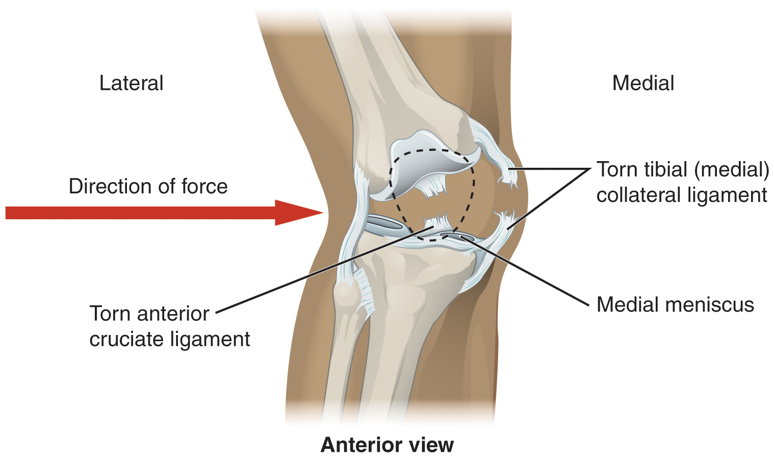
A worse combination of injuries can occur with a hit to the lateral side of the extended knee ( [link] ). A moderate blow to the lateral knee will cause the medial side of the joint to open, resulting in stretching or damage to the tibial collateral ligament. Because the medial meniscus is attached to the tibial collateral ligament, a stronger blow can tear the ligament and also damage the medial meniscus. This is one reason that the medial meniscus is 20 times more likely to be injured than the lateral meniscus. A powerful blow to the lateral knee produces a “terrible triad” injury, in which there is a sequential injury to the tibial collateral ligament, medial meniscus, and anterior cruciate ligament.
Arthroscopic surgery has greatly improved the surgical treatment of knee injuries and reduced subsequent recovery times. This procedure involves a small incision and the insertion into the joint of an arthroscope, a pencil-thin instrument that allows for visualization of the joint interior. Small surgical instruments are also inserted via additional incisions. These tools allow a surgeon to remove or repair a torn meniscus or to reconstruct a ruptured cruciate ligament. The current method for anterior cruciate ligament replacement involves using a portion of the patellar ligament. Holes are drilled into the cruciate ligament attachment points on the tibia and femur, and the patellar ligament graft, with small areas of attached bone still intact at each end, is inserted into these holes. The bone-to-bone sites at each end of the graft heal rapidly and strongly, thus enabling a rapid recovery.
Notification Switch
Would you like to follow the 'Anatomy & Physiology: support and movement' conversation and receive update notifications?

|
|

|
|

|
|

|
|

|
|

|
|
|
|
|
|
|
|

|
|

|