<< Chapter < Page Chapter >> Page >
The semipermeable membrane of a cell is shown, with different concentrations of potassium cations, sodium cations, and chloride anions inside and outside the cell. The ions are represented by small, colored circles. In its resting state, the cell membrane is permeable to potassium and chloride ions, but it is impermeable to sodium ions. By diffusion, potassium cations travel out of the cell, going through the cell membrane and forming a layer of positive charge on the outer surface of the membrane. By diffusion, chloride anions go into the cell, going through the cell membrane and forming a layer of negative charge on the inner surface of the membrane. As a result, a voltage is set up across the cell membrane. The Coulomb force prevents all the ions from crossing the membrane.
The semipermeable membrane of a cell has different concentrations of ions inside and out. Diffusion moves the K + size 12{"K" rSup { size 8{+{}} } } {} and Cl - size 12{"Cl" rSup { size 8{ +- {}} } } {} ions in the direction shown, until the Coulomb force halts further transfer. This results in a layer of positive charge on the outside, a layer of negative charge on the inside, and thus a voltage across the cell membrane. The membrane is normally impermeable to Na + size 12{"Na" rSup { size 8{+{}} } } {} .
This is a graphical representation of a pulse of voltage, or action potential, inside a nerve cell. The voltage in millivolts is plotted along the vertical axis and the time in milliseconds is plotted along the horizontal axis. Initially, between zero and about two point eight milliseconds, the voltage is a constant at about minus ninety millivolts, corresponding to the resting state. Above this section of the graph, a window shows a small cross-section of the cell membrane, with a positively charged outer surface, a negatively charged inner surface, and no ions moving across the membrane. Between two point eight and four point two milliseconds, the voltage increases to a peak of fifty millivolts, corresponding to depolarization of the membrane. A window above this section shows sodium cations crossing the membrane, from outside to inside the cell, so that the membrane’s inner surface acquires a positive charge and its outer surface has a negative charge. Between about four point two and about five point five milliseconds, the voltage drops to a low of about minus one hundred and ten millivolts, corresponding to repolarization of the membrane. A window above this section shows potassium cations crossing the membrane, from inside to outside the cell, so that the membrane’s outer surface again acquires a positive charge and its inner surface has a negative charge. After that, the voltage rises slightly, going back to a constant of about minus ninety millivolts, corresponding to the resting state. This movement of sodium and potassium ions across the membrane is called active transport, and long-term active transport is shown in a window above the final part of the curve.
An action potential is the pulse of voltage inside a nerve cell graphed here. It is caused by movements of ions across the cell membrane as shown. Depolarization occurs when a stimulus makes the membrane permeable to Na + size 12{"Na" rSup { size 8{+{}} } } {} ions. Repolarization follows as the membrane again becomes impermeable to Na + , size 12{"Na" rSup { size 8{+{}} } } {} and K + size 12{"K" rSup { size 8{+{}} } } {} moves from high to low concentration. In the long term, active transport slowly maintains the concentration differences, but the cell may fire hundreds of times in rapid succession without seriously depleting them.

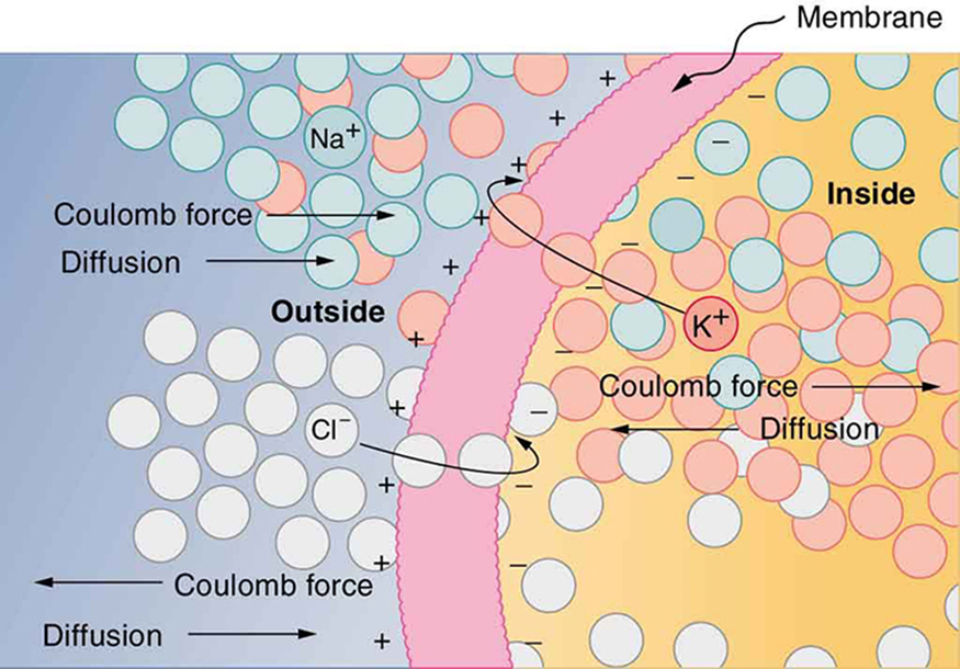
The separation of charge creates a potential difference of 70 to 90 mV across the cell membrane. While this is a small voltage, the resulting electric field ( E = V / d size 12{E = V/d} {} ) across the only 8-nm-thick membrane is immense (on the order of 11 MV/m!) and has fundamental effects on its structure and permeability. Now, if the exterior of a neuron is taken to be at 0 V, then the interior has a resting potential of about –90 mV. Such voltages are created across the membranes of almost all types of animal cells but are largest in nerve and muscle cells. In fact, fully 25% of the energy used by cells goes toward creating and maintaining these potentials.

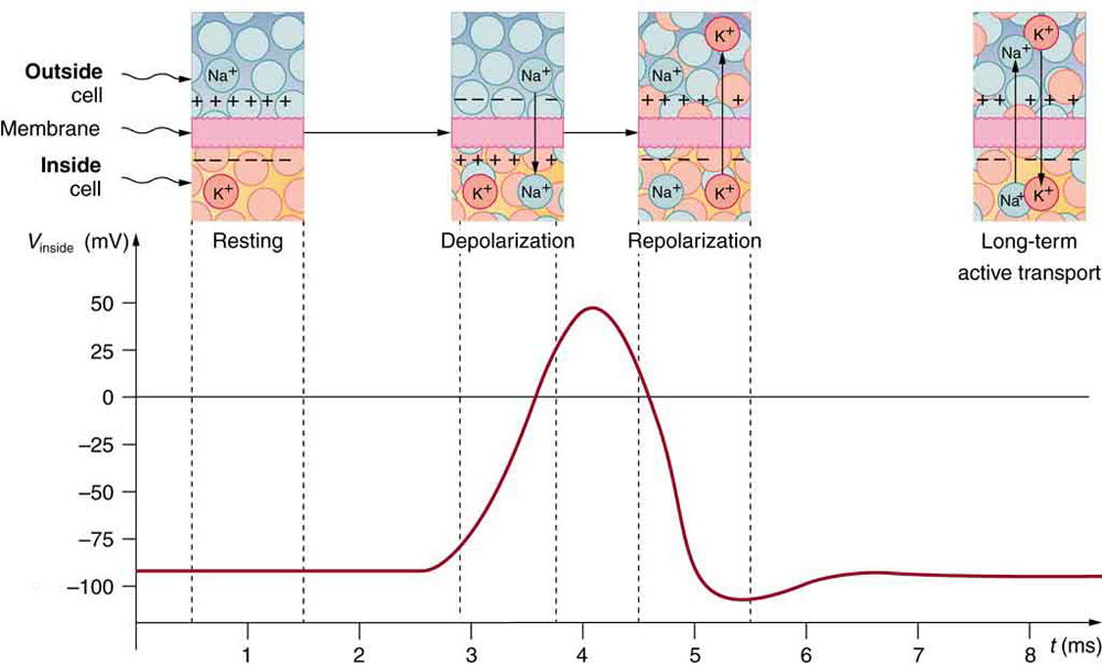
Electric currents along the cell membrane are created by any stimulus that changes the membrane’s permeability. The membrane thus temporarily becomes permeable to Na + size 12{"Na" rSup { size 8{+{}} } } {} , which then rushes in, driven both by diffusion and the Coulomb force. This inrush of Na + size 12{"Na" rSup { size 8{+{}} } } {} first neutralizes the inside membrane, or depolarizes it, and then makes it slightly positive. The depolarization causes the membrane to again become impermeable to Na + size 12{"Na" rSup { size 8{+{}} } } {} , and the movement of K + size 12{"K" rSup { size 8{+{}} } } {} quickly returns the cell to its resting potential, or repolarizes it. This sequence of events results in a voltage pulse, called the action potential . (See [link] .) Only small fractions of the ions move, so that the cell can fire many hundreds of times without depleting the excess concentrations of Na + size 12{"Na" rSup { size 8{+{}} } } {} and K + size 12{"K" rSup { size 8{+{}} } } {} . Eventually, the cell must replenish these ions to maintain the concentration differences that create bioelectricity. This sodium-potassium pump is an example of active transport , wherein cell energy is used to move ions across membranes against diffusion gradients and the Coulomb force.

The action potential is a voltage pulse at one location on a cell membrane. How does it get transmitted along the cell membrane, and in particular down an axon, as a nerve impulse? The answer is that the changing voltage and electric fields affect the permeability of the adjacent cell membrane, so that the same process takes place there. The adjacent membrane depolarizes, affecting the membrane further down, and so on, as illustrated in [link] . Thus the action potential stimulated at one location triggers a nerve impulse that moves slowly (about 1 m/s) along the cell membrane.

Practice Key Terms 4

Get Jobilize Job Search Mobile App in your pocket Now!

Get it on Google Play Download on the App Store Now


Source:  OpenStax, College physics ii. OpenStax CNX. Nov 29, 2012 Download for free at http://legacy.cnx.org/content/col11458/1.2
Google Play and the Google Play logo are trademarks of Google Inc.

Notification Switch

Would you like to follow the 'College physics ii' conversation and receive update notifications?

Ask
Edgar Delgado
Start Test
Anindyo Mukhopadhyay
Start Quiz
Rylee Minllic
Start Quiz
Madison Christian
Start Exam
Edgar Delgado
Start Quiz
Hope Percle
Start Quiz