<< Chapter < Page Chapter >> Page >
The illustration shows the tracheal system of a bee. Openings called spiracles appear along the side of the body. Vertical tubes lead from the spiracles to a tube that runs along the top of the body from front to back.
Insects perform respiration via a tracheal system.

Mammalian systems

In mammals, pulmonary ventilation occurs via inhalation (breathing). During inhalation, air enters the human body through the nasal cavity located just inside the nose ( [link] ). As air passes through the nasal cavity, the air is warmed to body temperature and humidified. The respiratory tract is coated with mucus to seal the tissues from direct contact with air. Mucus is high in water. As air crosses these surfaces of the mucous membranes, it picks up water. These processes help equilibrate the air to the body conditions, reducing any damage that cold, dry air can cause. Particulate matter that is floating in the air is removed in the nasal passages via mucus and cilia. The processes of warming, humidifying, and removing particles are important protective mechanisms that prevent damage to the trachea and lungs. Thus, inhalation serves several purposes in addition to bringing oxygen into the respiratory system.

The illustration shows the flow of air through the human respiratory system. The nasal cavity is a wide cavity above and behind the nostrils, and the pharynx is the passageway behind the mouth. The nasal cavity and pharynx join and enter the trachea through the larynx. The larynx is somewhat wider than the trachea and flat. The trachea has concentric, ring-like grooves, giving it a bumpy appearance. The trachea bifurcates into two primary bronchi, which are also grooved. The primary bronchi enter the lungs, and branch into secondary bronchi. The secondary bronchi in turn branch into many tertiary bronchi. The tertiary bronchi branch into bronchioles, which branch into terminal bronchioles. Each terminal bronchiole ends in an alveolar sac. Each alveolar sac contains many alveoli clustered together, like bunches of grapes. The alveolar duct is the air passage into the alveolar sac. The alveoli are hollow, and air empties into them. Pulmonary arteries bring deoxygenated blood to the alveolar sac (and thus appear blue), and pulmonary veins return oxygenated blood (and thus appear red) to the heart. Capillaries form a web around each alveolus. The diaphragm is a membrane that pushes up against the lungs.
Air enters the respiratory system through the nasal cavity and pharynx, and then passes through the trachea and into the bronchi, which bring air into the lungs. (credit: modification of work by NCI)

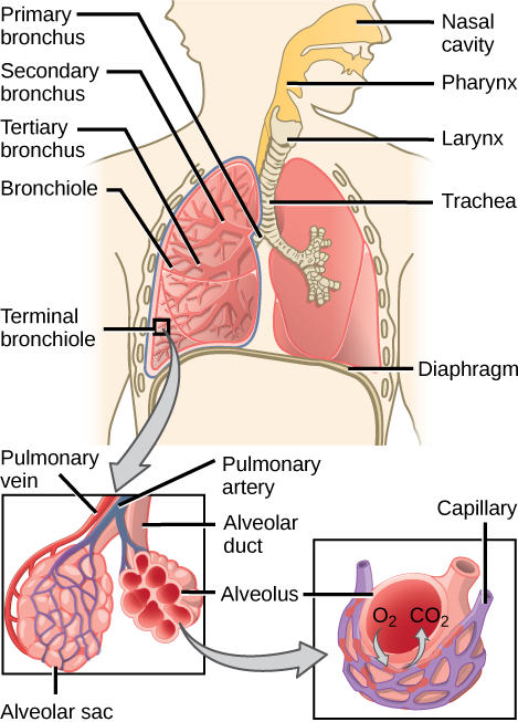
From the nasal cavity, air passes through the pharynx (throat) and the larynx (voice box), as it makes its way to the trachea ( [link] ). The main function of the trachea is to funnel the inhaled air to the lungs and the exhaled air back out of the body. The human trachea is a cylinder about 10 to 12 cm long and 2 cm in diameter that sits in front of the esophagus and extends from the larynx into the chest cavity where it divides into the two primary bronchi at the midthorax ( [link] ). The trachea is lined with mucus-producing goblet cells and ciliated epithelia. The cilia propel foreign particles trapped in the mucus toward the pharynx. The cartilage provides strength and support to the trachea to keep the passage open. The smooth muscle can contract, decreasing the trachea’s diameter, which causes expired air to rush upwards from the lungs at a great force. The forced exhalation helps expel mucus when we cough. Smooth muscle can contract or relax, depending on stimuli from the external environment or the body’s nervous system.

Lungs: bronchi and alveoli

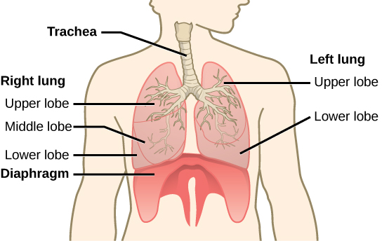
The end of the trachea bifurcates (divides) to the right and left lungs. The lungs are not identical. The right lung is larger and contains three lobes, whereas the smaller left lung contains two lobes ( [link] ). The muscular diaphragm , which facilitates breathing, is inferior (below) to the lungs and marks the end of the thoracic cavity.

The illustration shows the trachea, which starts at the top of the neck and continues down into the chest, where it branches into the bronchi, which enter the lungs. The left lung has two lobes. The upper lobe is located in front of and above the lower lobe. The right lung has three lobes. The upper lobe is on the top, the lower lobe is on the bottom, and the middle lobe is sandwiched between them. The diaphragm presses against the bottom of the lungs and has the appearance of skin stretched over the top of a drum. Wide flaps of the diaphragm extend downward on the front left and right sides of the body. On the back, thin flaps of diaphragm stretch downward on either side of the spine.
The trachea bifurcates into the right and left bronchi in the lungs. The right lung is made of three lobes and is larger. To accommodate the heart, the left lung is smaller and has only two lobes.

In the lungs, air is diverted into smaller and smaller passages, or bronchi. Air enters the lungs through the two primary (main) bronchi (singular: bronchus). Each bronchus divides into secondary bronchi, then into tertiary bronchi, which in turn divide, creating smaller and smaller diameter bronchioles as they split and spread through the lung.

Get Jobilize Job Search Mobile App in your pocket Now!

Get it on Google Play Download on the App Store Now


Source:  OpenStax, Principles of biology. OpenStax CNX. Aug 09, 2016 Download for free at http://legacy.cnx.org/content/col11569/1.25
Google Play and the Google Play logo are trademarks of Google Inc.

Notification Switch

Would you like to follow the 'Principles of biology' conversation and receive update notifications?

Ask