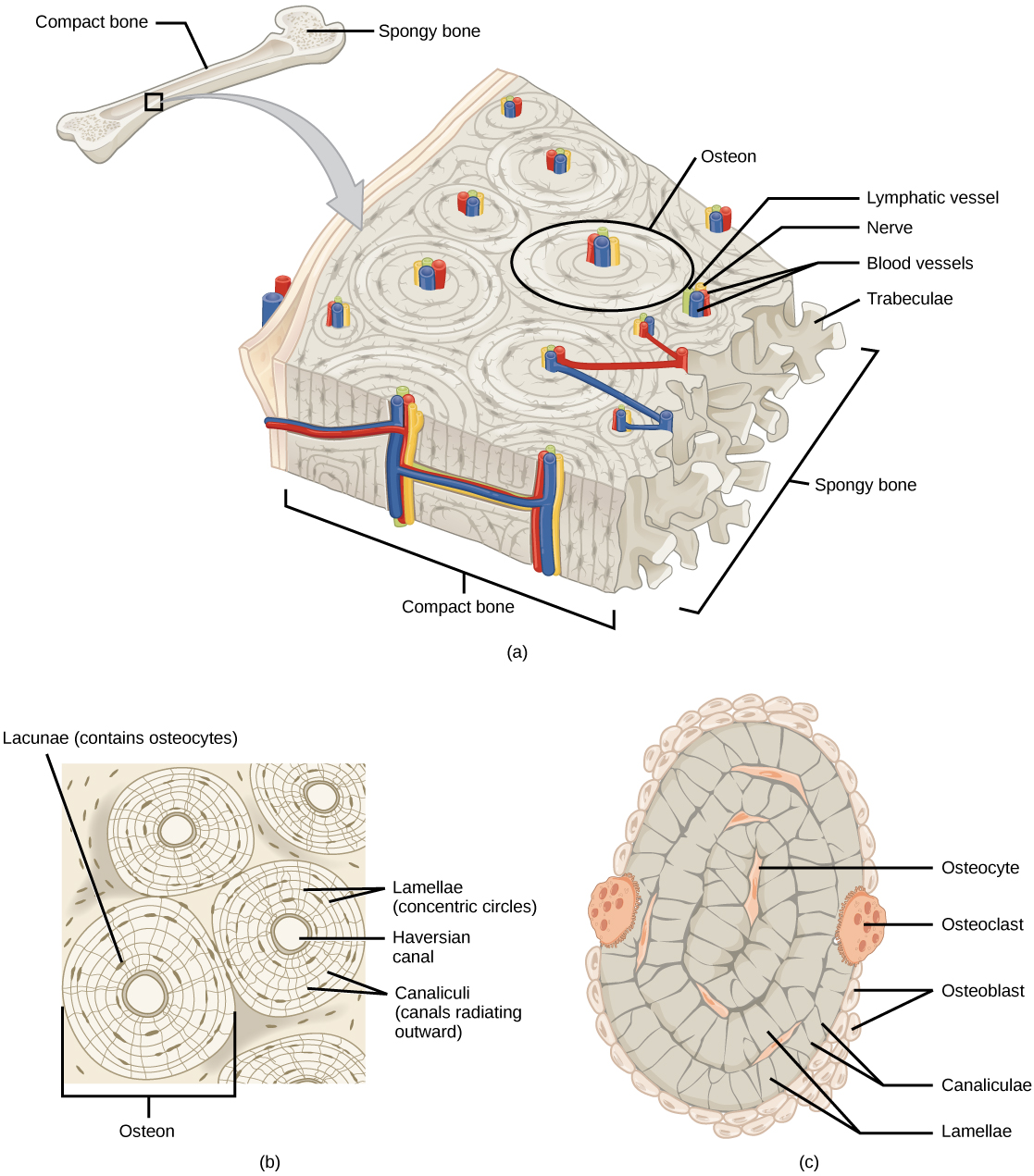
<< Chapter < Page Chapter >> Page >
 Illustration A shows a cross section of a long bone with wide protrusions at either end. The outer part is compact bone. Inside the compact bone is porous spongy bone made of web-like trabreculae. The spongy bone fills the wide part at either end of the bone. In the middle, a hollow exists inside the spongy bone. Illustration B shows several circular osteons clustered together in compact bone. At the hub of each osteon is an opening called the Haversian canal filled with blood and lymph vessels and nerves. The lamellae surrounding the Haversian canal resemble tree rings.  Lacunae are wide spaces in the rings between the lamellae. Microchannels called canaliculi radiate through the rings out from the central Haversian canal, connecting the lacunae together. Illustration C shows small osteoclasts surrounding the outside of bone. Larger osteoclasts are also on the outer surface, forming a hollow in the bone. Osteocytes are long, thin cells in the lacunae.
(a) Compact bone is a dense matrix on the outer surface of bone. Spongy bone, inside the compact bone, is porous with web-like trabeculae. (b) Compact bone is organized into rings called osteons. Blood vessels, nerves, and lymphatic vessels are found in the central Haversian canal. Rings of lamellae surround the Haversian canal. Between the lamellae are cavities called lacunae. Canaliculi are microchannels connecting the lacunae together. (c) Osteoblasts surround the exterior of the bone. Osteoclasts bore tunnels into the bone and osteocytes are found in the lacunae.

Adipose tissue

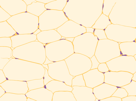
Adipose tissue, or fat tissue, is considered a connective tissue even though it does not have fibroblasts or a real matrix, and only has a few fibers. Adipose tissue is made up of cells called adipocytes (descendants of fibroblasts) that collect and store fat in the form of triglycerides. Adipose tissues serve as energy stores, and additionally serve as insulation to help maintain body temperatures, allowing animals to be endothermic. They also function as cushions to prevent damage to body organs. Under a microscope, adipose tissue cells appear empty due to the extraction of fat during the processing of the material for viewing, as seen in [link] . The thin lines in the image are the cell membranes, and the nuclei are the small, black dots at the edges of the cells.

Illustration shows irregularly shaped cells with tiny nuclei clustered next to the cell’s outer membrane.
Adipose is a connective tissue is made up of cells called adipocytes. Adipocytes have small nuclei localized at the cell edge. The interior of these large cells is filled with triglycerides, commonly known as "fat".

Blood

Blood is considered a connective tissue because it has a fluid matrix (plasma) and is derived from the germ layer known as mesoderm. . The living cell types are red blood cells (RBC), also called erythrocytes, and white blood cells (WBC), also called leukocytes ( [link] ).

Different types of blood cells are shown. Red blood cells are disc-shaped, with a central indentation. Platelets are much smaller than red blood cells, narrow and long. Neutrophils, eosinophils, lymphocytes, monocytes and basophils are all about three times the diameter of a red blood cell and round. They differ in the shape of the nucleus, and in the presence or absence of granules in the cytoplasm. Macrophages, which are the largest cell type, have pseudopods which give them an irregular shape.
Blood is a connective tissue that has a fluid matrix, called plasma, and no fibers. Erythrocytes (red blood cells), the predominant cell type, are involved in the transport of oxygen and carbon dioxide. Also present are various leukocytes (white blood cells) involved in immune response.

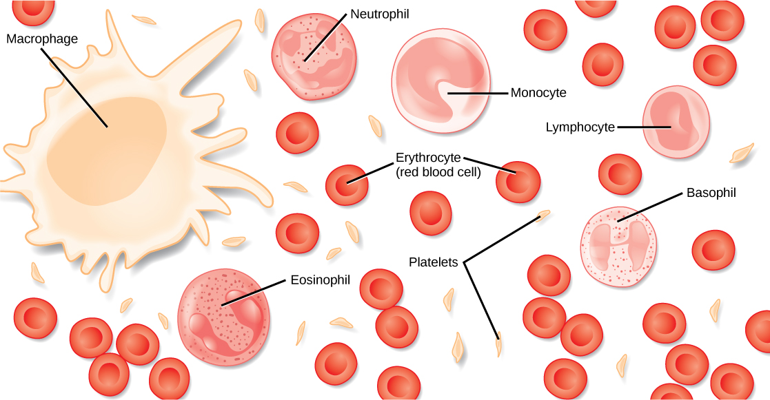
The cell found in greatest abundance in blood is the erythrocyte, or red blood cell ("erythro" = red). The principal function of an erythrocyte is to carry and deliver oxygen to the tissues. There are millions of erythrocytes in every milliliter of your blood. Mammalian erythrocytes lose their nuclei and mitochondria when they mature and are released from the bone marrow where they are generated. Fish, amphibian, and avian red blood cells maintain their nuclei and mitochondria throughout the cell’s life.

Leukocytes, or white blood cells ("leuko" = white), are the other main cellular component of blood. There are 5,000-10,000 leukocytes in every milliliter of your blood. These include cells called lymphocytes, as well as neutrophils, monocytes, and others. Lymphocytes function primarily in the immune response to foreign antigens or material, which you will learn in more detail later in this unit. Neutrophils are phagocytic (they engulf other cells or objects and digest them) cells, and they participate in one of the early lines of defense against microbial invaders or fungal invaders. Another leukocyte that is found in the peripheral blood is the monocyte. Monocytes give rise to phagocytic macrophages that clean up dead and damaged cells in the body, whether they are foreign or from the host animal. Two additional leukocytes in the blood are eosinophils and basophils—both help to facilitate the inflammatory response.

Get Jobilize Job Search Mobile App in your pocket Now!

Get it on Google Play Download on the App Store Now


Source:  OpenStax, Principles of biology. OpenStax CNX. Aug 09, 2016 Download for free at http://legacy.cnx.org/content/col11569/1.25
Google Play and the Google Play logo are trademarks of Google Inc.

Notification Switch

Would you like to follow the 'Principles of biology' conversation and receive update notifications?

Ask