<< Chapter < Page Chapter >> Page >
Part a shows red algae with lettuce-like leaves. Part b shows four oval green algae cells stacked next to each other. The cyanobacteria are about 2 µm across and 10 µm long.
(a) Red algae and (b) green algae (visualized by light microscopy) share similar DNA sequences with photosynthetic cyanobacteria. Scientists speculate that, in a process called endosymbiosis, an ancestral prokaryote engulfed a photosynthetic cyanobacterium that evolved into modern-day chloroplasts. (credit a: modification of work by Ed Bierman; credit b: modification of work by G. Fahnenstiel, NOAA; scale-bar data from Matt Russell)

Art connection

The illustration shows steps that, according to the endosymbiotic theory, gave rise to eukaryotic organisms. In step 1, infoldings in the plasma membrane of an ancestral prokaryote gave rise to endomembrane components, including a nucleus and endoplasmic reticulum. In step 2, the first endosymbiotic event occurred: The ancestral eukaryote consumed aerobic bacteria that evolved into mitochondria. In a second endosymbiotic event, the early eukaryote consumed photosynthetic bacteria that evolved into chloroplasts.
The first eukaryote may have originated from an ancestral prokaryote that had undergone membrane proliferation, compartmentalization of cellular function (into a nucleus, lysosomes, and an endoplasmic reticulum), and the establishment of endosymbiotic relationships with an aerobic prokaryote, and, in some cases, a photosynthetic prokaryote, to form mitochondria and chloroplasts, respectively.

What evidence is there that mitochondria were incorporated into the ancestral eukaryotic cell before chloroplasts?

Evolution connection

Secondary endosymbiosis in chlorarachniophytes

Endosymbiosis involves one cell engulfing another to produce, over time, a coevolved relationship in which neither cell could survive alone. The chloroplasts of red and green algae, for instance, are derived from the engulfment of a photosynthetic cyanobacterium by an early prokaryote.

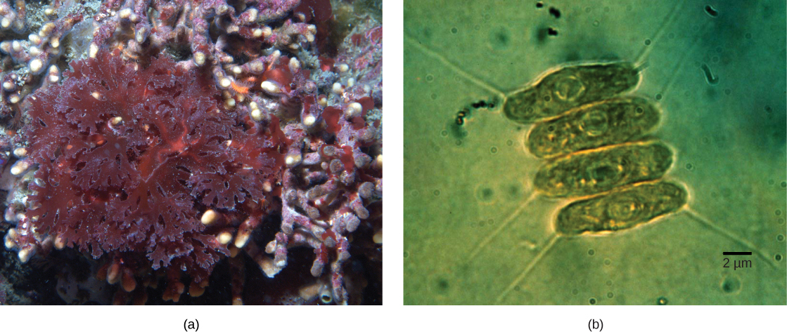
This leads to the question of the possibility of a cell containing an endosymbiont to itself become engulfed, resulting in a secondary endosymbiosis. Molecular and morphological evidence suggest that the chlorarachniophyte protists are derived from a secondary endosymbiotic event. Chlorarachniophytes are rare algae indigenous to tropical seas and sand that can be classified into the rhizarian supergroup. Chlorarachniophytes extend thin cytoplasmic strands, interconnecting themselves with other chlorarachniophytes, in a cytoplasmic network. These protists are thought to have originated when a eukaryote engulfed a green alga, the latter of which had already established an endosymbiotic relationship with a photosynthetic cyanobacterium ( [link] ).

According to the secondary endosymbiosis theory, plastids in modern chlorarachniophytes arose via two endosymbiotic events. In the first event, a cyanobacterium was engulfed by a heterotrophic eukaryote. Cyanobacteria have two membranes and the endosymbiosis event gave rise to a third membrane. One of these membranes was lost. Then, in a second endosymbiotic event, the cell was engulfed by another cell. The first cell became a plastid, an organelle with a vestigial nucleus and an organelle membrane inside it; thus, the plastid has the appearance of a cell within a cell.
The hypothesized process of endosymbiotic events leading to the evolution of chlorarachniophytes is shown. In a primary endosymbiotic event, a heterotrophic eukaryote consumed a cyanobacterium. In a secondary endosymbiotic event, the cell resulting from primary endosymbiosis was consumed by a second cell. The resulting organelle became a plastid in modern chlorarachniophytes.

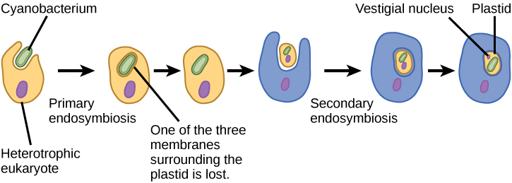
Several lines of evidence support that chlorarachniophytes evolved from secondary endosymbiosis. The chloroplasts contained within the green algal endosymbionts still are capable of photosynthesis, making chlorarachniophytes photosynthetic. The green algal endosymbiont also exhibits a stunted vestigial nucleus. In fact, it appears that chlorarachniophytes are the products of an evolutionarily recent secondary endosymbiotic event. The plastids of chlorarachniophytes are surrounded by four membranes: The first two correspond to the inner and outer membranes of the photosynthetic cyanobacterium, the third corresponds to the green alga, and the fourth corresponds to the vacuole that surrounded the green alga when it was engulfed by the chlorarachniophyte ancestor. In other lineages that involved secondary endosymbiosis, only three membranes can be identified around plastids. This is currently rectified as a sequential loss of a membrane during the course of evolution.

The process of secondary endosymbiosis is not unique to chlorarachniophytes. In fact, secondary endosymbiosis of green algae also led to euglenid protists, whereas secondary endosymbiosis of red algae led to the evolution of dinoflagellates, apicomplexans, and stramenopiles.

Section summary

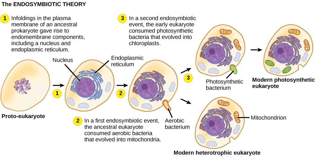
The oldest fossil evidence of eukaryotes is about 2 billion years old. Fossils older than this all appear to be prokaryotes. It is probable that today’s eukaryotes are descended from an ancestor that had a prokaryotic organization. The last common ancestor of today’s Eukarya had several characteristics, including cells with nuclei that divided mitotically and contained linear chromosomes where the DNA was associated with histones, a cytoskeleton and endomembrane system, and the ability to make cilia/flagella during at least part of its life cycle. It was aerobic because it had mitochondria that were the result of an aerobic alpha-proteobacterium that lived inside a host cell. Whether this host had a nucleus at the time of the initial symbiosis remains unknown. The last common ancestor may have had a cell wall for at least part of its life cycle, but more data are needed to confirm this hypothesis. Today’s eukaryotes are very diverse in their shapes, organization, life cycles, and number of cells per individual.

Art connections

[link] What evidence is there that mitochondria were incorporated into the ancestral eukaryotic cell before chloroplasts?

[link] All eukaryotic cells have mitochondria, but not all eukaryotic cells have chloroplasts.

Questions & Answers

what is biology
Hajah Reply
the study of living organisms and their interactions with one another and their environments
AI-Robot
what is biology
Victoria Reply
HOW CAN MAN ORGAN FUNCTION
Alfred Reply
the diagram of the digestive system
Assiatu Reply
allimentary cannel
Ogenrwot
How does twins formed
William Reply
They formed in two ways first when one sperm and one egg are splited by mitosis or two sperm and two eggs join together
Oluwatobi
what is genetics
Josephine Reply
Genetics is the study of heredity
Misack
how does twins formed?
Misack
What is manual
Hassan Reply
discuss biological phenomenon and provide pieces of evidence to show that it was responsible for the formation of eukaryotic organelles
Joseph Reply
what is biology
Yousuf Reply
the study of living organisms and their interactions with one another and their environment.
Wine
discuss the biological phenomenon and provide pieces of evidence to show that it was responsible for the formation of eukaryotic organelles in an essay form
Joseph Reply
what is the blood cells
Shaker Reply
list any five characteristics of the blood cells
Shaker
lack electricity and its more savely than electronic microscope because its naturally by using of light
Abdullahi Reply
advantage of electronic microscope is easily and clearly while disadvantage is dangerous because its electronic. advantage of light microscope is savely and naturally by sun while disadvantage is not easily,means its not sharp and not clear
Abdullahi
cell theory state that every organisms composed of one or more cell,cell is the basic unit of life
Abdullahi
is like gone fail us
DENG
cells is the basic structure and functions of all living things
Ramadan
What is classification
ISCONT Reply
is organisms that are similar into groups called tara
Yamosa
in what situation (s) would be the use of a scanning electron microscope be ideal and why?
Kenna Reply
A scanning electron microscope (SEM) is ideal for situations requiring high-resolution imaging of surfaces. It is commonly used in materials science, biology, and geology to examine the topography and composition of samples at a nanoscale level. SEM is particularly useful for studying fine details,
Hilary
cell is the building block of life.
Condoleezza Reply
Got questions? Join the online conversation and get instant answers!
Jobilize.com Reply

Get Jobilize Job Search Mobile App in your pocket Now!

Get it on Google Play Download on the App Store Now


Source:  OpenStax, Ucd bis2a intro to biology v1.2. OpenStax CNX. Sep 22, 2015 Download for free at https://legacy.cnx.org/content/col11890/1.1
Google Play and the Google Play logo are trademarks of Google Inc.

Notification Switch

Would you like to follow the 'Ucd bis2a intro to biology v1.2' conversation and receive update notifications?

Ask