<< Chapter < Page Chapter >> Page >
Chlamydia, Spirochetes, Cyanobacteria, and Gram-positive bacteria are described in this table. Note that bacterial shape is not phylum-dependent; bacteria within a phylum may be cocci, rod-shaped, or spiral. (credit “Chlamydia trachomatis”: modification of work by Dr. Lance Liotta Laboratory, NCI; credit “Treponema pallidum”: modification of work by Dr. David Cox, CDC; credit “Phormidium”: modification of work by USGS; credit “Clostridium difficile”: modification of work by Lois S. Wiggs, CDC; scale-bar data from Matt Russell)
Characteristics of the four phyla of archaea are described. Euryarchaeotes includes methanogens, which produce methane as a metabolic waste product, and halobacteria, which live in an extreme saline environment. Methanogens cause flatulence in humans and other animals. Halobacteria can grow in large blooms that appear reddish, due to the presence of bacterirhodopsin in the membrane. Bacteriorhodopsin is related to the retinal pigment rhodopsin. Micrograph shows rod-shaped Halobacterium. Members of the ubiquitous Crenarchaeotes phylum play an important role in the fixation of carbon. Many members of this group are sulfur-dependent extremophiles. Some are thermophilic or hyperthermophilic. Micrograph shows cocci-shaped Sulfolobus, a genus which grows in volcanic springs at temperatures between 75° and 80°C and at a pH between 2 and 3. The phylum Nanoarchaeotes currently contains only one species, Nanoarchaeum equitans, which has been isolated from the bottom of the Atlantic Ocean, and from the a hydrothermal vent at Yellowstone National Park. It is an obligate symbiont with Ignococcus, another species of archaebacteria. Micrograph shows two small, round N. equitans cells attached to a larger Ignococcus cell. Korarchaeotes are considered to be one of the most primitive forms of life and so far have only been found in the Obsidian Pool, a hot spring at Yellowstone National Park. Micrograph shows a variety of specimens from this group which vary in shape.
Archaea are separated into four phyla: the Korarchaeota, Euryarchaeota, Crenarchaeota, and Nanoarchaeota. (credit “Halobacterium”: modification of work by NASA; credit “Nanoarchaeotum equitans”: modification of work by Karl O. Stetter; credit “korarchaeota”: modification of work by Office of Science of the U.S. Dept. of Energy; scale-bar data from Matt Russell)

The plasma membrane

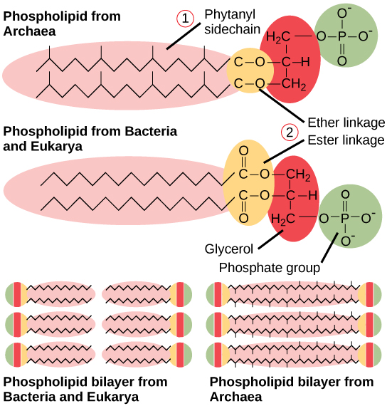
The plasma membrane is a thin lipid bilayer (6 to 8 nanometers) that completely surrounds the cell and separates the inside from the outside. Its selectively permeable nature keeps ions, proteins, and other molecules within the cell and prevents them from diffusing into the extracellular environment, while other molecules may move through the membrane. Recall that the general structure of a cell membrane is a phospholipid bilayer composed of two layers of lipid molecules. In archaeal cell membranes, isoprene (phytanyl) chains linked to glycerol replace the fatty acids linked to glycerol in bacterial membranes. Some archaeal membranes are lipid monolayers instead of bilayers ( [link] ).

This illustration compares phospholipids from Bacteria and Eukarya to those from Archaea. In Bacteria and Eukarya, fatty acids are attached to glycerol by an ester linkage, while in Archaea, isoprene chains are linked to glycerol by an ether linkage. In the ester linkage, the first carbon in the fatty acid chain has an oxygen double-bonded to it, whereas in the ether linkage, it does not. In Archaea, the isoprene chains have methyl groups branching off from them, whereas such branches are absent in Bacteria and Eukarya.  Both types of phospholipids result in similar lipid bilayers.
Archaeal phospholipids differ from those found in Bacteria and Eukarya in two ways. First, they have branched phytanyl sidechains instead of linear ones. Second, an ether bond instead of an ester bond connects the lipid to the glycerol.

The cell wall

The cytoplasm of prokaryotic cells has a high concentration of dissolved solutes. Therefore, the osmotic pressure within the cell is relatively high. The cell wall is a protective layer that surrounds some cells and gives them shape and rigidity. It is located outside the cell membrane and prevents osmotic lysis (bursting due to increasing volume). The chemical composition of the cell walls varies between archaea and bacteria, and also varies between bacterial species.

Bacterial cell walls contain peptidoglycan    , composed of polysaccharide chains that are cross-linked by unusual peptides containing both L- and D-amino acids including D-glutamic acid and D-alanine. Proteins normally have only L-amino acids; as a consequence, many of our antibiotics work by mimicking D-amino acids and therefore have specific effects on bacterial cell wall development. There are more than 100 different forms of peptidoglycan. S-layer    (surface layer) proteins are also present on the outside of cell walls of both archaea and bacteria.

Bacteria are divided into two major groups: Gram positive    and Gram negative    , based on their reaction to Gram staining. Note that all Gram-positive bacteria belong to one phylum; bacteria in the other phyla (Proteobacteria, Chlamydias, Spirochetes, Cyanobacteria, and others) are Gram-negative. The Gram staining method is named after its inventor, Danish scientist Hans Christian Gram (1853–1938). The different bacterial responses to the staining procedure are ultimately due to cell wall structure. Gram-positive organisms typically lack the outer membrane found in Gram-negative organisms ( [link] ). Up to 90 percent of the cell wall in Gram-positive bacteria is composed of peptidoglycan, and most of the rest is composed of acidic substances called teichoic acids . Teichoic acids may be covalently linked to lipids in the plasma membrane to form lipoteichoic acids. Lipoteichoic acids anchor the cell wall to the cell membrane. Gram-negative bacteria have a relatively thin cell wall composed of a few layers of peptidoglycan (only 10 percent of the total cell wall), surrounded by an outer envelope containing lipopolysaccharides (LPS) and lipoproteins. This outer envelope is sometimes referred to as a second lipid bilayer. The chemistry of this outer envelope is very different, however, from that of the typical lipid bilayer that forms plasma membranes.

Questions & Answers

Discuss the differences between taste and flavor, including how other sensory inputs contribute to our  perception of flavor.
John Reply
taste refers to your understanding of the flavor . while flavor one The other hand is refers to sort of just a blend things.
Faith
While taste primarily relies on our taste buds, flavor involves a complex interplay between taste and aroma
Kamara
which drugs can we use for ulcers
Ummi Reply
omeprazole
Kamara
what
Renee
what is this
Renee
is a drug
Kamara
of anti-ulcer
Kamara
Omeprazole Cimetidine / Tagament For the complicated once ulcer - kit
Patrick
what is the function of lymphatic system
Nency Reply
Not really sure
Eli
to drain extracellular fluid all over the body.
asegid
The lymphatic system plays several crucial roles in the human body, functioning as a key component of the immune system and contributing to the maintenance of fluid balance. Its main functions include: 1. Immune Response: The lymphatic system produces and transports lymphocytes, which are a type of
asegid
to transport fluids fats proteins and lymphocytes to the blood stream as lymph
Adama
what is anatomy
Oyindarmola Reply
Anatomy is the identification and description of the structures of living things
Kamara
what's the difference between anatomy and physiology
Oyerinde Reply
Anatomy is the study of the structure of the body, while physiology is the study of the function of the body. Anatomy looks at the body's organs and systems, while physiology looks at how those organs and systems work together to keep the body functioning.
AI-Robot
what is enzymes all about?
Mohammed Reply
Enzymes are proteins that help speed up chemical reactions in our bodies. Enzymes are essential for digestion, liver function and much more. Too much or too little of a certain enzyme can cause health problems
Kamara
yes
Prince
how does the stomach protect itself from the damaging effects of HCl
Wulku Reply
little girl okay how does the stomach protect itself from the damaging effect of HCL
Wulku
it is because of the enzyme that the stomach produce that help the stomach from the damaging effect of HCL
Kamara
function of digestive system
Ali Reply
function of digestive
Ali
the diagram of the lungs
Adaeze Reply
what is the normal body temperature
Diya Reply
37 degrees selcius
Xolo
37°c
Stephanie
please why 37 degree selcius normal temperature
Mark
36.5
Simon
37°c
Iyogho
the normal temperature is 37°c or 98.6 °Fahrenheit is important for maintaining the homeostasis in the body the body regular this temperature through the process called thermoregulation which involves brain skin muscle and other organ working together to maintain stable internal temperature
Stephanie
37A c
Wulku
what is anaemia
Diya Reply
anaemia is the decrease in RBC count hemoglobin count and PVC count
Eniola
what is the pH of the vagina
Diya Reply
how does Lysin attack pathogens
Diya
acid
Mary
I information on anatomy position and digestive system and there enzyme
Elisha Reply
anatomy of the female external genitalia
Muhammad Reply
Organ Systems Of The Human Body (Continued) Organ Systems Of The Human Body (Continued)
Theophilus Reply
what's lochia albra
Kizito
Got questions? Join the online conversation and get instant answers!
Jobilize.com Reply

Get Jobilize Job Search Mobile App in your pocket Now!

Get it on Google Play Download on the App Store Now


Source:  OpenStax, Ucd bis2a intro to biology v1.2. OpenStax CNX. Sep 22, 2015 Download for free at https://legacy.cnx.org/content/col11890/1.1
Google Play and the Google Play logo are trademarks of Google Inc.

Notification Switch

Would you like to follow the 'Ucd bis2a intro to biology v1.2' conversation and receive update notifications?

Ask