

| << Chapter < Page | Chapter >> Page > |
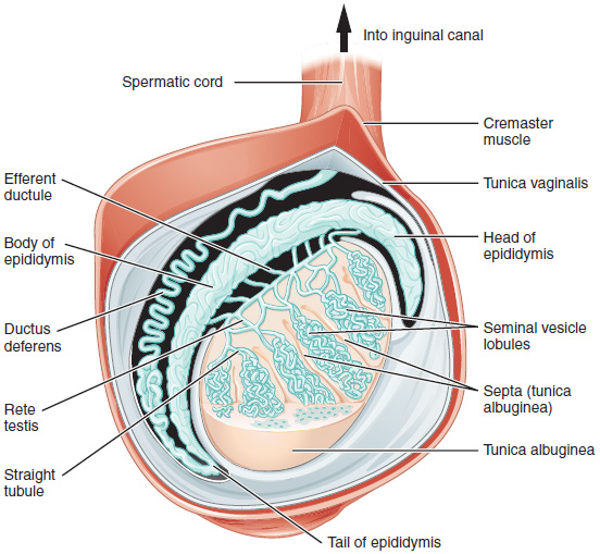
The tightly coiled seminiferous tubules form the bulk of each testis. They are where sperm are formed and released into the duct system of the testis. Specifically, from the seminiferous tubules, sperm move into the straight tubules (or tubuli recti), and from there into a fine meshwork of tubules called the rete testes . Sperm leave the rete testes, and the testis itself, through the 15 to 20 efferent ductules of the testes.
Surrounding all stages of the developing sperm cells are elongate, branching Sertoli cells . Sertoli cells are a type of supporting cell. Sertoli cells secrete signaling molecules that promote sperm production.
The least mature cells, the spermatogonia (singular = spermatogonium), line the inside the tubule. Spermatogonia are the stem cells of the testis, which means that they are still able to differentiate into a variety of different cell types throughout adulthood. Spermatogonia divide to produce primary and secondary spermatocytes , then spermatids , which finally produce formed sperm. The process that begins with spermatogonia and concludes with the production of sperm is called spermatogenesis .
The process of spermatogenesis begins at puberty, after which time sperm are produced constantly throughout a man’s life. One production cycle, from spermatogonia through formed sperm, takes approximately 64 days. Sperm counts —the total number of sperm a man produces—slowly decline after age 35.
The process of spermatogenesis begins with mitosis of the diploid spermatogonia ( [link] ). However, mature gametes are haploid (1 n ), containing 23 chromosomes—meaning that the spermatogonia must undergo a second cellular division through the process of meiosis .
Start second reading here*************************** .
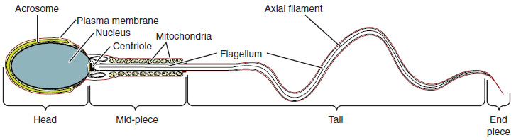
Sperm are smaller than most cells in the body; in fact, the volume of a sperm cell is 85,000 times less than that of the female gamete. Approximately 100 to 300 million sperm are produced each day, whereas women typically ovulate only one oocyte per month. The structure of sperm cells speaks to their function. Sperm have a distinctive head, mid-piece, and tail region ( [link] ). The head of the sperm contains a nucleus with very little cytoplasm. A structure called the acrosome covers most of the head of the sperm cell as a “cap” that is filled with enzymes important for preparing sperm to participate in fertilization. Tightly packed mitochondria fill the mid-piece of the sperm. ATP produced by these mitochondria will power the flagellum , which extends from the neck and the mid-piece through the tail of the sperm, enabling it to move the entire sperm cell.
Notification Switch
Would you like to follow the 'Mrs browne's reproductive modules' conversation and receive update notifications?

|
|

|
|

|
|
|
|
|

|
|

|
|
|
|

|
|
|
|
|

|