<< Chapter < Page Chapter >> Page >

Thin skin versus thick skin

Part A is a micrograph showing a cross section of thin skin. The topmost layer is a thin, translucent layer with irregular texture and areas where cells are sloughing off. The deepest layer is dark purple and extends into the third layer with finger like projections. The third light purple layer contains thin bands of fibers and small, dark cells. The fourth, and deepest layer, is darker than the third layer, but is still light purple. It contains thick fiber bands that are loosely packed. Part B is a magnified view of the epidermis of thick skin. It shows the topmost layer is five times thicker than the topmost layer of thin skin. The topmost layer of thick skin is also denser and less translucent than the topmost layer of thin skin.
These slides show cross-sections of the epidermis and dermis of (a) thin and (b) thick skin. Note the significant difference in the thickness of the epithelial layer of the thick skin. From top, LM × 40, LM × 40. (Micrographs provided by the Regents of University of Michigan Medical School © 2012)

The cells in all of the layers except the stratum basale are called keratinocytes. A keratinocyte    is a cell that manufactures and stores the protein keratin. Keratin is an intracellular fibrous protein that gives hair, nails, and skin their hardness and water-resistant properties. The keratinocytes in the stratum corneum are dead and regularly slough away, being replaced by cells from the deeper layers ( [link] ).

Epidermis

The outer layer of cells in this micrograph is the thinnest layer and stained deep purple due to full keratinization of dead cells. The next layer occupies one quarter of the micrograph, is lightly stained, and is a dense collection of cells. The third layer from the top is mostly white, with lightly stained, loosely-packed strands radiating in random directions. The bottom-most layer is densely-packed, with thick bands of highly organized muscle tissue that are darkly stained.
The epidermis is epithelium composed of multiple layers of cells. The basal layer consists of cuboidal cells, whereas the outer layers are squamous, keratinized cells, so the whole epithelium is often described as being keratinized stratified squamous epithelium. LM × 40. (Micrograph provided by the Regents of University of Michigan Medical School © 2012)

Stratum basale

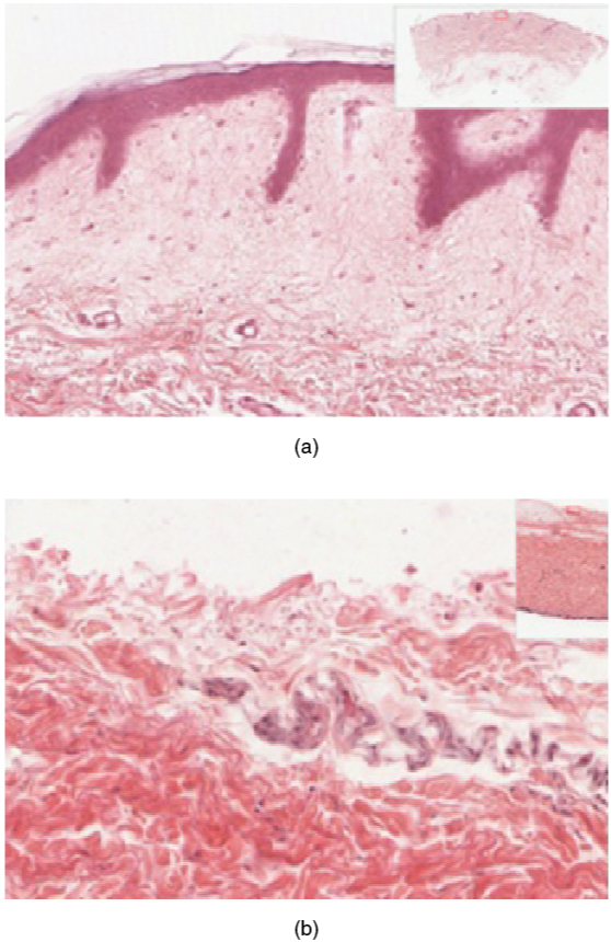
The stratum basale    (also called the stratum germinativum) is the deepest epidermal layer and attaches the epidermis to the the connective tissue of the dermis. The cells in the stratum basale bond to the dermis via intertwining collagen fibers, referred to as the basement membrane. A finger-like projection, or fold, known as the dermal papilla    (plural = dermal papillae) is found in the superficial portion of the dermis. Dermal papillae increase the strength of the connection between the epidermis and dermis; the greater the folding, the stronger the connections made ( [link] ).

Layers of the epidermis

This illustration shows a cross section of the epidermis. The cells of the innermost layer, the stratum basale, are large and have a purple nucleus. The stratum basale curls around the dermis, which projects into the epidermis. The stratum basale contains four layers of large, triangle-shaped keratinocytes. Fibers are visible within the spaces between keratinocytes in the stratum basale. A melanocyte is also present in this layer. The melanocyte possesses finger-like projections extending from its main cell body. The projections branch through the extracellular spaces between nearby keratinocytes. Above the stratum basale is the stratum spinosum which consists of 8 layers of oval-shaped keratinocytes. The nucleus is present in these keratinocytes, but has faded to a lighter purple. The stratum granulosum contains five layers of keratinocytes, each containing spots in its cytoplasm, labeled the lamellar granules. The stratum lucidium contains 4 layers of diamond-shaped cells with no nucleus. The stratum corneum contains 9 layers of keratinocytes with no nucleus , nor cytoplasm. A few of the cells in the topmost layer of the stratum corneum are flaking off from the skin.
The epidermis of thick skin has five layers: stratum basale, stratum spinosum, stratum granulosum, stratum lucidum, and stratum corneum.

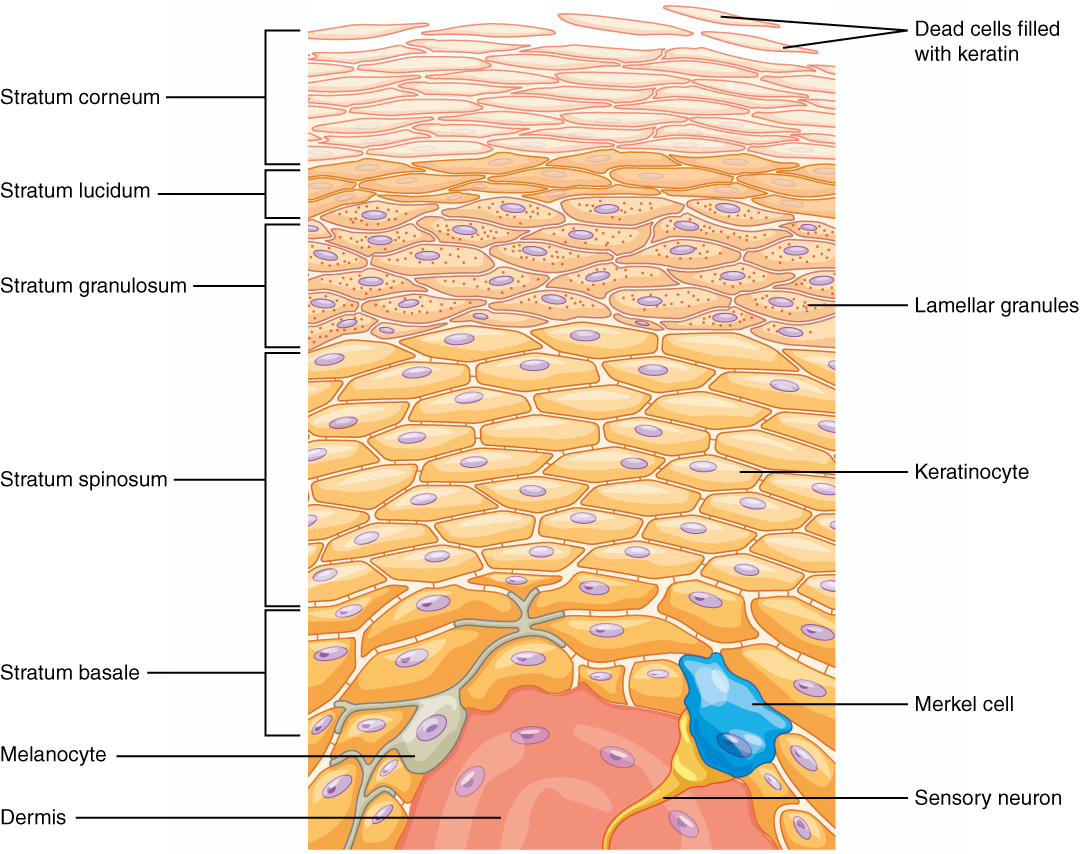
The stratum basale is a single layer of cells primarily made of basal cells. A basal cell    which forms keratinocytes of the epidermis. All of the keratinocytes are produced from this single layer of cells, which are constantly going through mitosis to produce new cells. As new cells are formed, the existing cells are pushed superficially away from the stratum basale. The second is a melanocyte    , a cell that produces the pigment melanin. Melanin gives hair and skin its color, and also helps protect the living cells of the epidermis from ultraviolet (UV) radiation damage.

In a growing fetus, fingerprints form where the cells of the stratum basale meet the papillae of the underlying dermal layer (papillary layer), resulting in the formation of the ridges on your fingers that you recognize as fingerprints. Fingerprints are unique to each individual and are used for forensic analyses because the patterns do not change with the growth and aging processes.

Stratum spinosum

As the name suggests, the stratum spinosum    is spiny in appearance due to the protruding cell processes that join the cells via a structure called a desmosome    . The desmosomes interlock with each other and strengthen the bond between the cells. The stratum spinosum is composed of eight to 10 layers of keratinocytes, formed as a result of cell division in the stratum basale ( [link] ).

Get Jobilize Job Search Mobile App in your pocket Now!

Get it on Google Play Download on the App Store Now


Source:  OpenStax, Integumentary system. OpenStax CNX. Mar 19, 2015 Download for free at http://legacy.cnx.org/content/col11770/1.1
Google Play and the Google Play logo are trademarks of Google Inc.

Notification Switch

Would you like to follow the 'Integumentary system' conversation and receive update notifications?

Ask