A schematic representation of coordination number in different layers in which there are two shells around the center atom. Both shells, green (x) and red (+), have coordination numbers of 4, but the radial distance of the red one (+) is bigger than the green one (x). Based on S. D. Kelly, D. Hesterberg, and B. Ravel in
Methods of Soil Analysis: Part 5, Mineralogical Methods , Ed. A. L. Urely and R. Drees, Soil Science Society of America Book Series, Madison (2008).
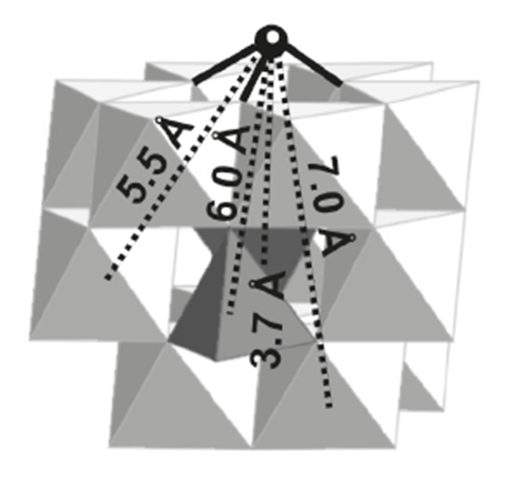
An EXAFS signal is given by the photoelectron scattering generated for the center atom. The phase of the signal is determinate by the distance and the path the photoelectrons travel. A simple scheme of the different paths is shown by
[link] . In the case of two shells around the centered atom, there is a degeneracy of four for the path between the main atom to the first shell, a degeneracy of four for the path between the main atom to the second shell, and a degeneracy of eight for the path between the main atom to the first shell, to the second one and to the center atom.
A two shell diagram in which there are three kinds of paths. From the center atom to the green one (x) and then going back (1); from the center atom to the red one (+) and the going back (2); and from the center atom to the first shell to the second one, and the returning to the center atom (3). Based on S. D. Kelly, D. Hesterberg, and B. Ravel in
Methods of Soil Analysis: Part 5, Mineralogical Methods , Ed. A. L. Urely and R. Drees, Soil Science Society of America Book Series, Madison (2008).
The analysis of EXAFS spectra is accomplished using Fourier transformation to fit the data to the EXAFS equation. The EXAFS equation is a sum of the contribution from all scattering paths of the photoelectrons
[link] , where each path is given by
[link] .
The terms
Feffi (k), φ
i (k), and
λi (k) are the effective scattering amplitude of the photoelectron, the phase shift of the photoelectron, and the mean free path of the photoelectron, respectively. The term
R
i is the half path length of the photoelectron (the distance between the centered atom and a coordinating atom for a single-scattering event). And the
k2 is given by the
[link] . The remaining variables are frequently determined by modeling the EXAFS spectrum.
Xafs analysis for arsenic adsorption onto iron oxides
The absorption of arsenic species onto iron oxide offers n example of the information that can be obtained by EXAFS. Because the huge impact that the presence of arsenic in water can produce in societies there is a lot of research in the adsorption of arsenic in several kinds of materials, in particular nano materials. Some of the materials more promising for this kind of applications are iron oxides. The elucidation of the mechanism of arsenic coordination onto the surfaces of those materials has been studied lately using X-ray absorption spectroscopy.
There are several ways how arsenate (AsO
43− ,
[link] ) can be adsorbed onto the surfaces.
[link] shows the three ways that Sherman proposes arsenate can be adsorbed onto goethite (α-FeOOH): bidentate cornersharing (2C), bidentate edge sharing (2E) and monodentate corner-sharing (1V) shapes.
[link] shows that the bidentate corner sharing (2C) is the configuration that corresponds with the calculated parameters not only for goethite, but for several iron oxides.
Structure of the arsenate anion.Possible configurations of arsenate onto goethite. The tetrahedral with the small spheres represents the arsenate ions. Adapted from D. M. Sherman and S. R. Randal.
Geochim. Cosmochim. Ac. 2003,
67 , 4223.Fourier transforms of the EXAFS for arsenate sorbed onto goethite, lepidocrocite, hematite and ferrihydrite. Adapted from D. M. Sherman and S. R. Randal.
Geochim. Cosmochim. Ac. 2003,
67 , 4223.
Several studies have confirmed that the bidentate corner sharing (2C) is the one present in the arsenate adsorption but also one similar, a tridentate corner sharing complex (3C), for the arsenite adsorption onto most of iron oxides as shows
[link] .
[link] shows the coordination numbers and distances reported in the literature for the As(III) and As(V) onto goethite.
Proposed structural model for arsenic(III) tridante. Adapted from G. Morin, Y. Wang, G. Ona-Nguema, F. Juillot, G. Calas, N. Menguy, E. Aubry, J. R. Bargar, and G. E. Brown.
Langmuir 2009,
25 , 9119.
As
CNAs-O
RAs-O(Å)
CNAs-Fe
RAs-Fe(Å)
III
3.06±0.03
1.79±0.8
2.57±0.01
3.34±3
3.19
1.77±1
1.4
3.34±5
3
1.78
2
3.55±5
V
1.03.0
1.631.70
2
3.30
4.6
1.68
--
3.55±5
Coordination numbers (CN) and inter-atomic distances (R) reported in the literature for the As(III) and As(V) adsorption onto goethite.
Bibliography
G. Bunker.
Introduction to XAFS: A practical guide to X-ray Absorption Fine Structure Spectroscopy , Cambridge University Press, Cambridge (2010).
S. D. Kelly, D. Hesterberg, and B. Ravel in
Methods of Soil Analysis: Part 5, Mineralogical Methods , Ed. A. L. Urely and R. Drees, Soil Science Society of America Book Series, Madison (2008).
G. A. Waychunas, B. A. Rea, C. C. Fuller, and J. A. Davis.
Geochim. Cosmochim. Ac. 1993,
57 , 2251.
B. A. Manning, and S. E. Fendorf, and S.
Goldberg
, Environ. Sci. Techn ., 1998,
32 , 2383.
D. M. Sherman and S. R. Randal,
Geochim. Cosmochim. Ac. , 2003,
67 , 4223.
G. Ona-Nguena, G. Morin F. Juillot, G. Calas, and G. E. Brown,
Environ. Sci. Techn. , 2005,
39 , 9147.
M. Stachowicz, T. Hiemstra, and W. H. Van Riemsdijk,
J. Colloid. Interf. Sci. , 2005,
302 , 62.
M. Auffan, J. Rose, O. Proux, D. Borschneck, A. Masion, P. Chaurand, J. L. Hazemann, C. Chaneac, J. P. Jolivet, M. R. Wiesner, A. Van Geen, and J. Y. Bottero,
Langmuir , 2008,
24 , 3215.
G. Ona-Nguena, G. Morin, Y. Wang, N. Menguy, F. Juillot, L, Olivi, G. Aquilanti, M. Abdelmoula, C. Ruby, J. R. Bargar, F. Guyot, G. Calas, and G. E. Brown, Jr.,
Geochim. Cosmochim. Ac. , 2009,
73 , 1359.
G. Morin, Y. Wang, G. Ona-Nguema, F. Juillot, G. Calas, N. Menguy, E. Aubry, J. R. Bargar, and G. E. Brown Jr.,
Langmuir , 2009,
25 , 9119.
J. Rose, M. M. Cortalezzi-Fidalgo, S. Moustier, C. Magnetto, C. D. Jones, A. R. Barron, M. R. Wiesner, and J.-Y. Bottero,
Chem. Mater. , 2002,
14 , 621.