<< Chapter < Page Chapter >> Page >
The TEM diagram show a high voltage wire attached to an electron gun which releases a beam of electrons. The electron beam passes by the first condenser lens (connected to a condenser aperture), then the second condenser lens (also connected to a condenser aperture), and then through the specimen on the specimen holder and air lock (which is also connected to an objective lens and aperture). Finally, the electron beam travels to the fluorescent screen and camera. The SEM begins with an electron gun that fires electron beams through an anode, through a condenser lens, through scanning coils and on to the sample on the stage. A backscatter electron detector detects electrons that travel directly back from the sample; secondary electron detectors detect electrons that travel to the sides.
These schematic illustrations compare the components of transmission electron microscopes and scanning electron microscopes.
Figure a shows A TEM micrograph with a clear background and a dark cell in the center. A double line outlines the edge of the cell and webs of material inside the cell are visible. Figure b shows an SEM micrograph that has large purple clusters on a green background with small holes. The three dimensionality of the purple clusters is apparent.
(a) This TEM image of cells in a biofilm shows well-defined internal structures of the cells because of varying levels of opacity in the specimen. (b) This color-enhanced SEM image of the bacterium Staphylococcus aureus illustrates the ability of scanning electron microscopy to render three-dimensional images of the surface structure of cells. (credit a: modification of work by American Society for Microbiology; credit b: modification of work by Centers for Disease Control and Prevention)
  • What are some advantages and disadvantages of electron microscopy, as opposed to light microscopy, for examining microbiological specimens?
  • What kinds of specimens are best examined using TEM? SEM?

Using microscopy to study biofilms

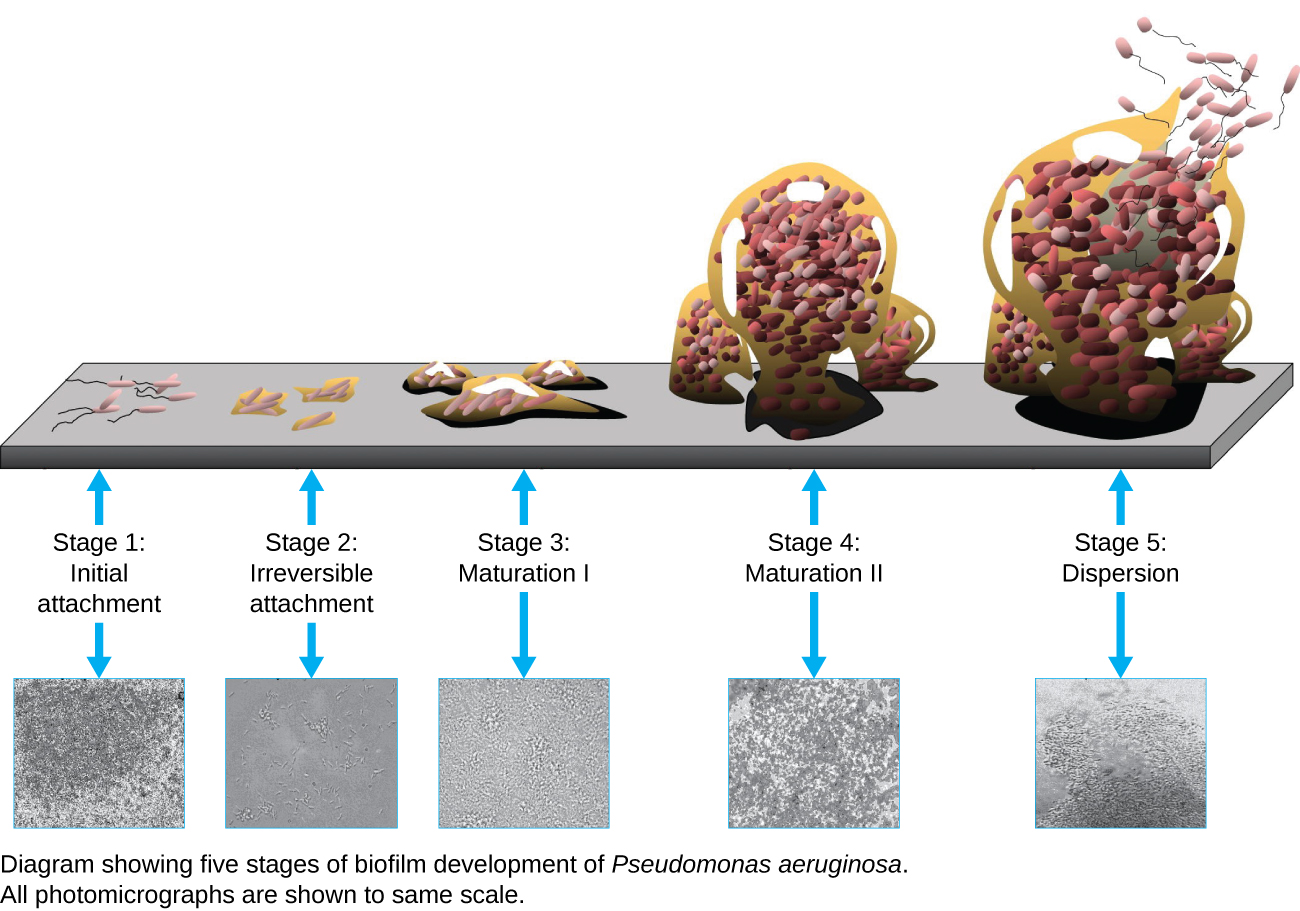
A biofilm is a complex community of one or more microorganism species, typically forming as a slimy coating attached to a surface because of the production of an extrapolymeric substance (EPS) that attaches to a surface or at the interface between surfaces (e.g., between air and water). In nature, biofilms are abundant and frequently occupy complex niches within ecosystems ( [link] ). In medicine, biofilms can coat medical devices and exist within the body. Because they possess unique characteristics, such as increased resistance against the immune system and to antimicrobial drugs, biofilms are of particular interest to microbiologists and clinicians alike.

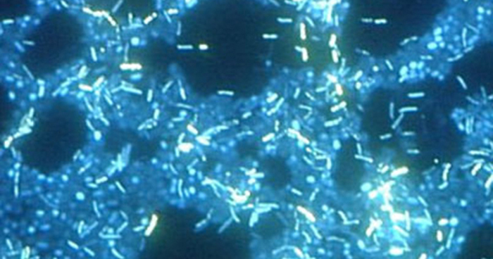
Because biofilms are thick, they cannot be observed very well using light microscopy; slicing a biofilm to create a thinner specimen might kill or disturb the microbial community. Confocal microscopy provides clearer images of biofilms because it can focus on one z-plane at a time and produce a three-dimensional image of a thick specimen. Fluorescent dyes can be helpful in identifying cells within the matrix. Additionally, techniques such as immunofluorescence and fluorescence in situ hybridization (FISH) , in which fluorescent probes are used to bind to DNA, can be used.

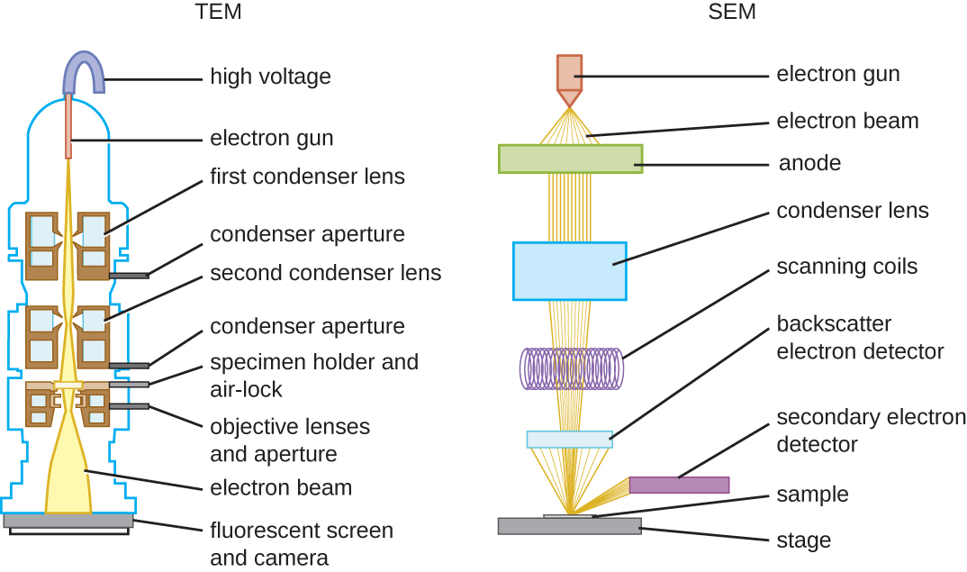
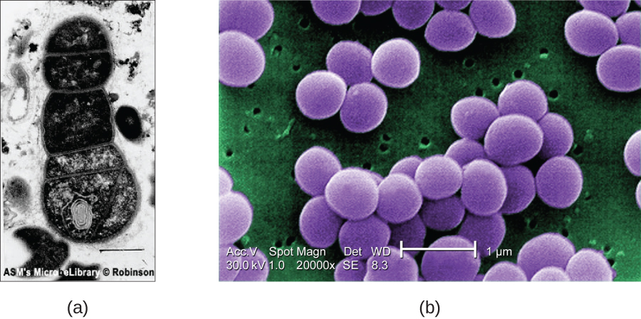
Electron microscopy can be used to observe biofilms, but only after dehydrating the specimen, which produces undesirable artifacts and distorts the specimen. In addition to these approaches, it is possible to follow water currents through the shapes (such as cones and mushrooms) of biofilms, using video of the movement of fluorescently coated beads ( [link] ).

The stages of a biofilm are shown. In stage 1 (initial attachment), a few flagellated cells attach to a surface. In stage 2 (irreversible attachment) clumps of cells are found on the surface. In stage 3 (maturation) the clumps have enlarged. In stage 4 (maturation 2) the clumps have fused and enlarged greatly. In stage 5 (dispersal) the large clump releases flagellated cells away from the surface. These stages are also shown in micrographs: 1) small dots, 2) larger clumps, 3)larger clump, 4) a large mass, 5) a large mass with an opening at the top.
A biofilm forms when planktonic (free-floating) bacteria of one or more species adhere to a surface, produce slime, and form a colony. (credit: Public Library of Science)
A micrograph with a black background containing many bright rectangles in clumps is shown.
In this image, multiple species of bacteria grow in a biofilm on stainless steel (stained with DAPI for epifluorescence miscroscopy). (credit: Ricardo Murga, Rodney Donlan)

Scanning probe microscopy

A scanning probe microscope does not use light or electrons, but rather very sharp probes that are passed over the surface of the specimen and interact with it directly. This produces information that can be assembled into images with magnifications up to 100,000,000⨯. Such large magnifications can be used to observe individual atoms on surfaces. To date, these techniques have been used primarily for research rather than for diagnostics.

Get Jobilize Job Search Mobile App in your pocket Now!

Get it on Google Play Download on the App Store Now


Source:  OpenStax, Microbiology. OpenStax CNX. Nov 01, 2016 Download for free at http://cnx.org/content/col12087/1.4
Google Play and the Google Play logo are trademarks of Google Inc.

Notification Switch

Would you like to follow the 'Microbiology' conversation and receive update notifications?

Ask