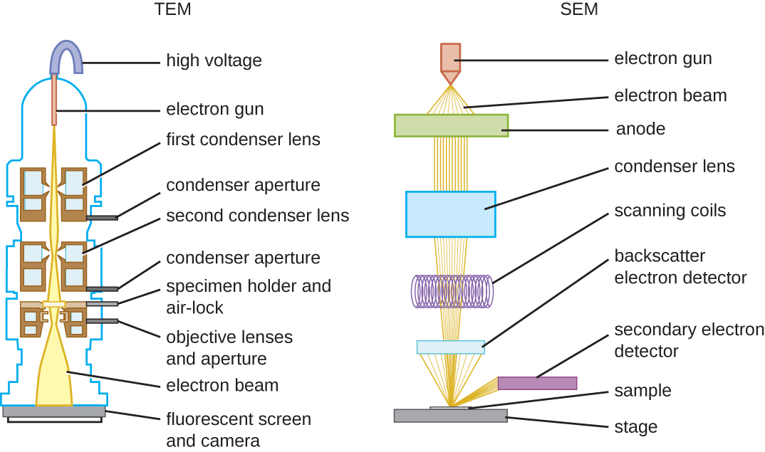
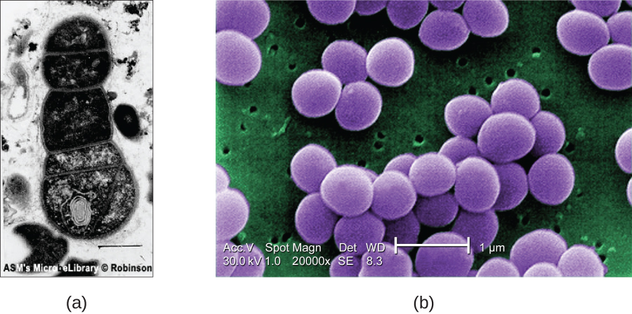
These schematic illustrations compare the components of transmission electron microscopes and scanning electron microscopes.(a) This TEM image of cells in a biofilm shows well-defined internal structures of the cells because of varying levels of opacity in the specimen. (b) This color-enhanced SEM image of the bacterium
Staphylococcus aureus illustrates the ability of scanning electron microscopy to render three-dimensional images of the surface structure of cells. (credit a: modification of work by American Society for Microbiology; credit b: modification of work by Centers for Disease Control and Prevention)
What are some advantages and disadvantages of electron microscopy, as opposed to light microscopy, for examining microbiological specimens?
What kinds of specimens are best examined using TEM? SEM?
Using microscopy to study biofilms
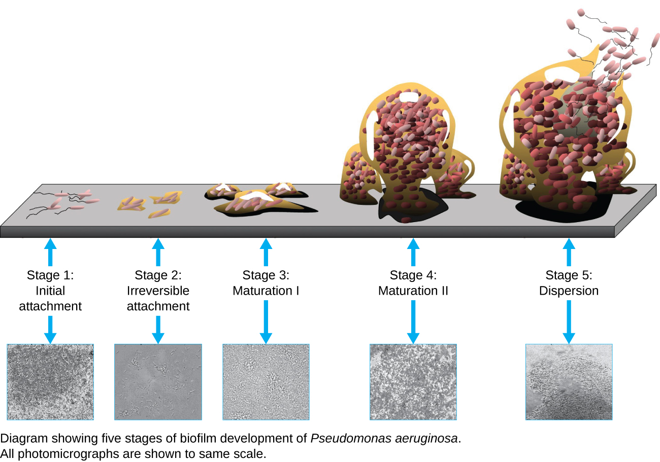
A biofilm is a complex community of one or more microorganism species, typically forming as a slimy coating attached to a surface because of the production of an
extrapolymeric substance (EPS) that attaches to a surface or at the interface between surfaces (e.g., between air and water). In nature, biofilms are abundant and frequently occupy complex niches within ecosystems (
[link] ). In medicine,
biofilms can coat medical devices and exist within the body. Because they possess unique characteristics, such as increased resistance against the immune system and to antimicrobial drugs, biofilms are of particular interest to microbiologists and clinicians alike.
Because biofilms are thick, they cannot be observed very well using light microscopy; slicing a biofilm to create a thinner specimen might kill or disturb the microbial community. Confocal microscopy provides clearer images of biofilms because it can focus on one z-plane at a time and produce a three-dimensional image of a thick specimen. Fluorescent dyes can be helpful in identifying cells within the matrix. Additionally, techniques such as immunofluorescence and
fluorescence in situ hybridization (FISH) , in which fluorescent probes are used to bind to DNA, can be used.
Electron microscopy can be used to observe biofilms, but only after dehydrating the specimen, which produces undesirable artifacts and distorts the specimen. In addition to these approaches, it is possible to follow water currents through the shapes (such as cones and mushrooms) of biofilms, using video of the movement of fluorescently coated beads (
[link] ).
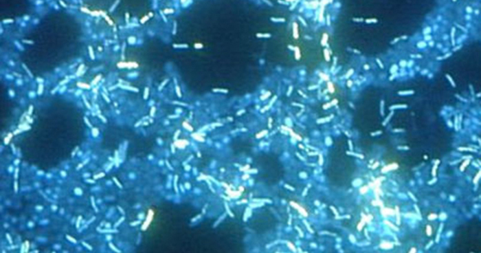
A biofilm forms when planktonic (free-floating) bacteria of one or more species adhere to a surface, produce slime, and form a colony. (credit: Public Library of Science)In this image, multiple species of bacteria grow in a biofilm on stainless steel (stained with DAPI for epifluorescence miscroscopy). (credit: Ricardo Murga, Rodney Donlan)
Scanning probe microscopy
A
scanning probe microscope does not use light or electrons, but rather very sharp probes that are passed over the surface of the specimen and interact with it directly. This produces information that can be assembled into images with magnifications up to 100,000,000⨯. Such large magnifications can be used to observe individual atoms on surfaces. To date, these techniques have been used primarily for research rather than for diagnostics.
Bacteria doesn't produce energy they are dependent upon their substrate in case of lack of nutrients they are able to make spores which helps them to sustain in harsh environments
_Adnan
But not all bacteria make spores, l mean Eukaryotic cells have Mitochondria which acts as powerhouse for them, since bacteria don't have it, what is the substitution for it?
Assimilatory nitrate reduction is a process that occurs in some microorganisms, such as bacteria and archaea, in which nitrate (NO3-) is reduced to nitrite (NO2-), and then further reduced to ammonia (NH3).
Elkana
This process is called assimilatory nitrate reduction because the nitrogen that is produced is incorporated in the cells of microorganisms where it can be used in the synthesis of amino acids and other nitrogen products
There are nothing like emergency disease but there are some common medical emergency which can occur simultaneously like Bleeding,heart attack,Breathing difficulties,severe pain heart stock.Hope you will get my point .Have a nice day ❣️
_Adnan
define infection ,prevention and control
Innocent
I think infection prevention and control is the avoidance of all things we do that gives out break of infections and promotion of health practices that promote life