

| << Chapter < Page | Chapter >> Page > |
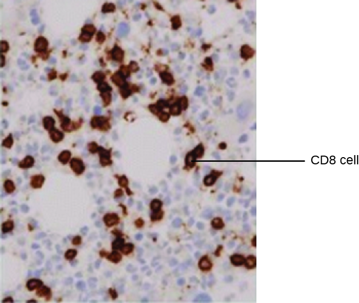
One powerful use of EIA is immunostaining , in which antibody-enzyme conjugates enhance microscopy. Immunohistochemistry (IHC) is used for examining whole tissues. As seen in [link] , a section of tissue can be stained to visualize the various cell types. In this example, a mAb against CD8 was used to stain CD8 cells in a section of tonsil tissue. It is now possible to count the number of CD8 cells, determine their relative numbers versus the other cell types present, and determine the location of these cells within this tissue. Such data would be useful for studying diseases such as AIDS, in which the normal function of CD8 cells is crucial for slowing disease progression.
Immunocytochemistry (ICC) is another valuable form of immunostaining. While similar to IHC, in ICC, extracellular matrix material is stripped away, and the cell membrane is etched with alcohol to make it permeable to antibodies. This allows antibodies to pass through the cell membrane and bind to specific targets inside the cell. Organelles, cytoskeletal components, and other intracellular structures can be visualized in this way. While some ICC techniques use EIA, the enzyme can be replaced with a fluorescent molecule, making it a fluorescent immunoassay.
The enzyme-linked immunosorbent assays (ELISAs) are widely used EIAs. In the direct ELISA , antigens are immobilized in the well of a microtiter plate. An antibody that is specific for a particular antigen and is conjugated to an enzyme is added to each well. If the antigen is present, then the antibody will bind. After washing to remove any unbound antibodies, a colorless substrate ( chromogen ) is added. The presence of the enzyme converts the substrate into a colored end product ( [link] ). While this technique is faster because it only requires the use of one antibody, it has the disadvantage that the signal from a direct ELISA is lower (lower sensitivity).
In a sandwich ELISA , the goal is to use antibodies to precisely quantify specific antigen present in a solution, such as antigen from a pathogen, a serum protein, or a hormone from the blood or urine to list just a few examples. The first step of a sandwich ELISA is to add the primary antibody to all the wells of a microtiter plate ( [link] ). The antibody sticks to the plastic by hydrophobic interactions. After an appropriate incubation time, any unbound antibody is washed away. Comparable washes are used between each of the subsequent steps to ensure that only specifically bound molecules remain attached to the plate. A blocking protein is then added (e.g., albumin or the milk protein casein) to bind the remaining nonspecific protein-binding sites in the well. Some of the wells will receive known amounts of antigen to allow the construction of a standard curve, and unknown antigen solutions are added to the other wells. The primary antibody captures the antigen and, following a wash, the secondary antibody is added, which is a polyclonal antibody that is conjugated to an enzyme. After a final wash, a colorless substrate (chromogen) is added, and the enzyme converts it into a colored end product. The color intensity of the sample caused by the end product is measured with a spectrophotometer . The amount of color produced (measured as absorbance) is directly proportional to the amount of enzyme, which in turn is directly proportional to the captured antigen. ELISAs are extremely sensitive, allowing antigen to be quantified in the nanogram (10 –9 g) per mL range.
Notification Switch
Would you like to follow the 'Microbiology' conversation and receive update notifications?

|
|

|
|

|
|

|
|

|
|
|
|
|

|
|

|
|

|
|
|
|