<< Chapter < Page Chapter >> Page >

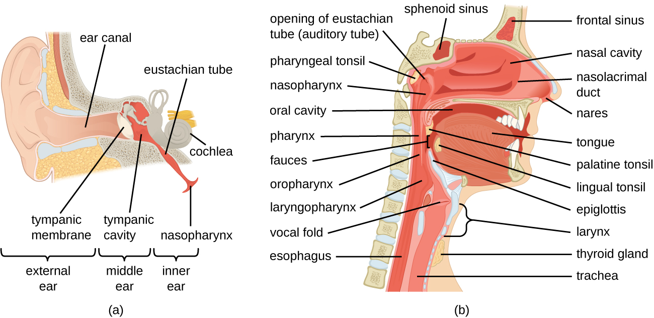
The oral cavity is a secondary opening for the respiratory tract. The oral and nasal cavities connect through the fauces to the pharynx, or throat. The pharynx can be divided into three regions: the nasopharynx, the oropharynx , and the laryngopharynx . Air inhaled through the mouth does not pass through the nasopharynx; it proceeds first through the oropharynx and then through the laryngopharynx. The palatine tonsils , which consist of lymphoid tissue, are located within the oropharynx. The laryngopharynx, the last portion of the pharynx, connects to the larynx , which contains the vocal fold ( [link] ).

a) diagram of ear; a closeup shows the bones and membranes of the middle ear. The eardrum is a flat disk labeled tympanic membrane. Behind this is the tympanic cavity (middle ear) which contains the bones. A tube boing downward from the middle ear is labeled Eustachian tube (auditory tube). b) A diagram of a cross section of the head. Above the nose is a space in the bone labeled frontal sinus.  The space in the nose is the nasal cavity and a duct in the nose is the nasolacrimal duct. A space in the bone behind the nose is the sphenoid sinus. At the back of the nose is the opening of the Eustachian tube (auditory tube). Behind that is the pharyngeal tonsil. Below that is a tube labeled nasopharynx which becomes the pharynx which because the oropharynx (behind the mouth) which becomes the laryngopharynx, which becomes the esophagus. Vocal folds are found just beyond the laryngopharynx in the larynx a tube which becomes the trachea. The epiglottis is a flap the determines if material in the pharynx travels to the esophagus or the trachea because the mouth also leads to the pharynx. The mouth contains the tongue. Underneath the tongue is the lingual tonsil and at the back of the mouth is the palatine tonsil. At the very back of the mouth is the fauces. In front of the trachea is the thyroid gland.
(a) The ear is connected to the upper respiratory tract by the eustachian tube, which opens to the nasopharynx. (b) The structures of the upper respiratory tract.
  • Identify the sequence of anatomical structures through which microbes would pass on their way from the nares to the larynx.
  • What two anatomical points do the eustachian tubes connect?

Anatomy of the lower respiratory system

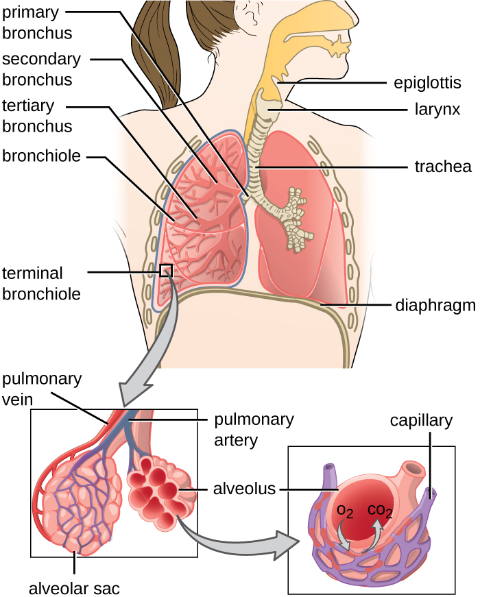
The lower respiratory system begins below the epiglottis in the larynx or voice box ( [link] ). The trachea , or windpipe, is a cartilaginous tube extending from the larynx that provides an unobstructed path for air to reach the lungs. The trachea bifurcates into the left and right bronchi as it reaches the lungs. These paths branch repeatedly to form smaller and more extensive networks of tubes, the bronchiole s. The terminal bronchioles formed in this tree-like network end in cul-de-sacs called the alveoli . These structures are surrounded by capillary networks and are the site of gas exchange in the respiratory system. Human lungs contain on the order of 400,000,000 alveoli. The outer surface of the lungs is protected with a double-layered pleural membrane. This structure protects the lungs and provides lubrication to permit the lungs to move easily during respiration.

A drawA drawing of the lower respiratory system. The epiglottis is a flap that can allow material into the larynx. The larynx is a tube that leads to the trachea. The trachea branches to become the primary bronchi. These branch to become the secondary bronchi, these branch to become the tertiary bronchi. These branch to become the bronchioles. Terminal bronchioles end in clusters of balloon shapes called alveolar sacs. Each balloon shape is an alveolus. Thin, webbed capillaries cover the outside of the alveolus and are connected to pulmonary veins and pulmonary arteries. Oxygen from the alveolus travels into the capillary and carbon dioxide from the capillary travels into the alveolus. ing of the lower respiratory system. The epiglottis is a flap that can allow material into the larynx. The larynx is a tube that leads to the trachea. The trachea branches to become the primary bronchi. These branch to become the secondary bronchi, these branch to become the tertiary bronchi. These branch to become the bronchioles. Terminal bronchioles end in clusters of balloon shapes called alveolar sacs. Each balloon shape is an alveolus. Thin, webbed capillaries cover the outside of the alveolus and are connected to pulmonary veins and pulmonary arteries. Oxygen from the alveolus travels into the capillary and carbon dioxide from the capillary travels into the alveolus.
The structures of the lower respiratory tract are identified in this illustration. (credit: modification of work by National Cancer Institute)

Defenses of the respiratory system

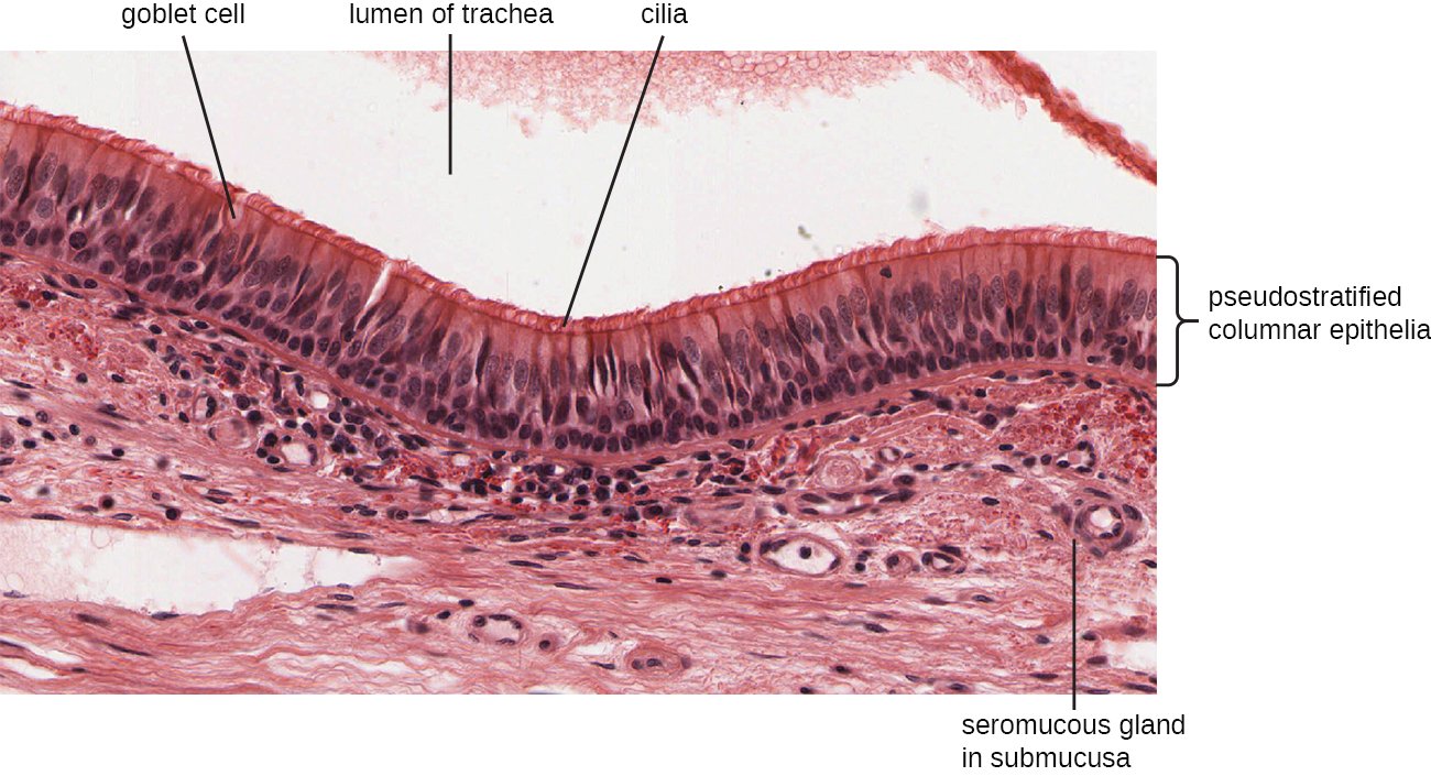
The inner lining of the respiratory system consists of mucous membrane s ( [link] ) and is protected by multiple immune defenses. The goblet cell s within the respiratory epithelium secrete a layer of sticky mucus. The viscosity and acidity of this secretion inhibits microbial attachment to the underlying cells. In addition, the respiratory tract contains ciliated epithelial cells. The beating cilia dislodge and propel the mucus, and any trapped microbes, upward to the epiglottis, where they will be swallowed. Elimination of microbes in this manner is referred to as the mucociliary escalator effect and is an important mechanism that prevents inhaled microorganisms from migrating further into the lower respiratory tract.

A micrograph showing a space at the top labeled lumen of trachea. Underneath this are long cells with a brush border at the top. These cells are called pseudostratified columnar epithelia. The brush border is many cilia. Vase shaped cells in this layer are called goblet cells. Below this layer is tissue with small spheres labeled seromucous gland in submucosa.
This micrograph shows the structure of the mucous membrane of the respiratory tract. (credit: modification of micrograph provided by the Regents of University of Michigan Medical School © 2012)

The upper respiratory system is under constant surveillance by mucosa-associated lymphoid tissue (MALT) , including the adenoids and tonsils. Other mucosal defenses include secreted antibodies (IgA), lysozyme, surfactant, and antimicrobial peptides called defensins . Meanwhile, the lower respiratory tract is protected by alveolar macrophage s. These phagocytes efficiently kill any microbes that manage to evade the other defenses. The combined action of these factors renders the lower respiratory tract nearly devoid of colonized microbes.

Questions & Answers

how does Neisseria cause meningitis
Nyibol Reply
what is microbiologist
Muhammad Reply
what is errata
Muhammad
is the branch of biology that deals with the study of microorganisms.
Ntefuni Reply
What is microbiology
Mercy Reply
studies of microbes
Louisiaste
when we takee the specimen which lumbar,spin,
Ziyad Reply
How bacteria create energy to survive?
Muhamad Reply
Bacteria doesn't produce energy they are dependent upon their substrate in case of lack of nutrients they are able to make spores which helps them to sustain in harsh environments
_Adnan
But not all bacteria make spores, l mean Eukaryotic cells have Mitochondria which acts as powerhouse for them, since bacteria don't have it, what is the substitution for it?
Muhamad
they make spores
Louisiaste
what is sporadic nd endemic, epidemic
Aminu Reply
the significance of food webs for disease transmission
Abreham
food webs brings about an infection as an individual depends on number of diseased foods or carriers dully.
Mark
explain assimilatory nitrate reduction
Esinniobiwa Reply
Assimilatory nitrate reduction is a process that occurs in some microorganisms, such as bacteria and archaea, in which nitrate (NO3-) is reduced to nitrite (NO2-), and then further reduced to ammonia (NH3).
Elkana
This process is called assimilatory nitrate reduction because the nitrogen that is produced is incorporated in the cells of microorganisms where it can be used in the synthesis of amino acids and other nitrogen products
Elkana
Examples of thermophilic organisms
Shu Reply
Give Examples of thermophilic organisms
Shu
advantages of normal Flora to the host
Micheal Reply
Prevent foreign microbes to the host
Abubakar
they provide healthier benefits to their hosts
ayesha
They are friends to host only when Host immune system is strong and become enemies when the host immune system is weakened . very bad relationship!
Mark
what is cell
faisal Reply
cell is the smallest unit of life
Fauziya
cell is the smallest unit of life
Akanni
ok
Innocent
cell is the structural and functional unit of life
Hasan
is the fundamental units of Life
Musa
what are emergency diseases
Micheal Reply
There are nothing like emergency disease but there are some common medical emergency which can occur simultaneously like Bleeding,heart attack,Breathing difficulties,severe pain heart stock.Hope you will get my point .Have a nice day ❣️
_Adnan
define infection ,prevention and control
Innocent
I think infection prevention and control is the avoidance of all things we do that gives out break of infections and promotion of health practices that promote life
Lubega
Heyy Lubega hussein where are u from?
_Adnan
en français
Adama
which site have a normal flora
ESTHER Reply
Many sites of the body have it Skin Nasal cavity Oral cavity Gastro intestinal tract
Safaa
skin
Asiina
skin,Oral,Nasal,GIt
Sadik
How can Commensal can Bacteria change into pathogen?
Sadik
How can Commensal Bacteria change into pathogen?
Sadik
all
Tesfaye
by fussion
Asiina
what are the advantages of normal Flora to the host
Micheal
what are the ways of control and prevention of nosocomial infection in the hospital
Micheal
what is inflammation
Shelly Reply
part of a tissue or an organ being wounded or bruised.
Wilfred
what term is used to name and classify microorganisms?
Micheal Reply
Binomial nomenclature
adeolu

Get Jobilize Job Search Mobile App in your pocket Now!

Get it on Google Play Download on the App Store Now


Source:  OpenStax, Microbiology. OpenStax CNX. Nov 01, 2016 Download for free at http://cnx.org/content/col12087/1.4
Google Play and the Google Play logo are trademarks of Google Inc.

Notification Switch

Would you like to follow the 'Microbiology' conversation and receive update notifications?

Ask