<< Chapter < Page Chapter >> Page >

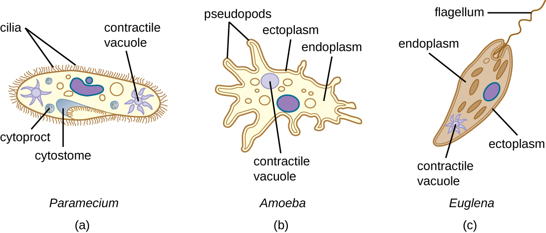
Many protists have whip-like flagella or hair-like cilia made of microtubules that can be used for locomotion ( [link] ). Other protists use cytoplasmic extensions known as pseudopodia (“false feet”) to attach the cell to a surface; they then allow cytoplasm to flow into the extension, thus moving themselves forward.

Protozoans have a variety of unique organelles and sometimes lack organelles found in other cells. Some have contractile vacuole s , organelles that can be used to move water out of the cell for osmotic regulation (salt and water balance) ( [link] ). Mitochondria may be absent in parasites or altered to kinetoplastids (modified mitochondria) or hydrogenosomes (see Unique Characteristics of Prokaryotic Cells for more discussion of these structures).

a) Paramecium cell with short strands on the outside labeled cilia. An indent in the outer layer is labeled cytostome. A sphere inside the cell at the base of the cytostome is labeled cytoproct. A star shaped structure inside the cell is labeled contractile vacuole. B) Amoeba cell with projections on the outside labeled pseudopods. The outer layer of the cell is labeled ectoplasm and the inner layer is labeled endoplasm. A sphere inside the cell is labeled contractile vacuole. C) Euglena with a single long flagellum on the outside. The outer layer of the cell is labeled etoplasm, the inner layer is labeled endoplasm. A star shaped structure is labeled contractile vacuole.
(a) Paramecium spp. have hair-like appendages called cilia for locomotion. (b) Amoeba spp. use lobe-like pseudopodia to anchor the cell to a solid surface and pull forward. (c) Euglena spp. use a whip-like structure called a flagellum to propel the cell.
  • What is the sequence of events in reproduction by schizogony and what are the cells produced called?

Taxonomy of protists

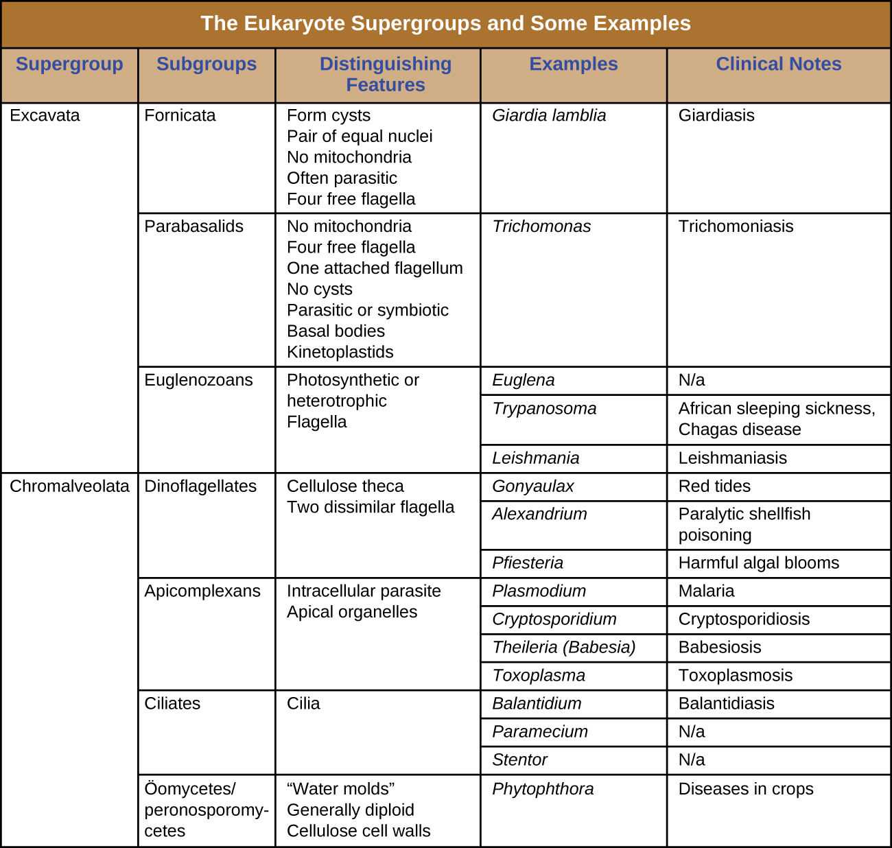
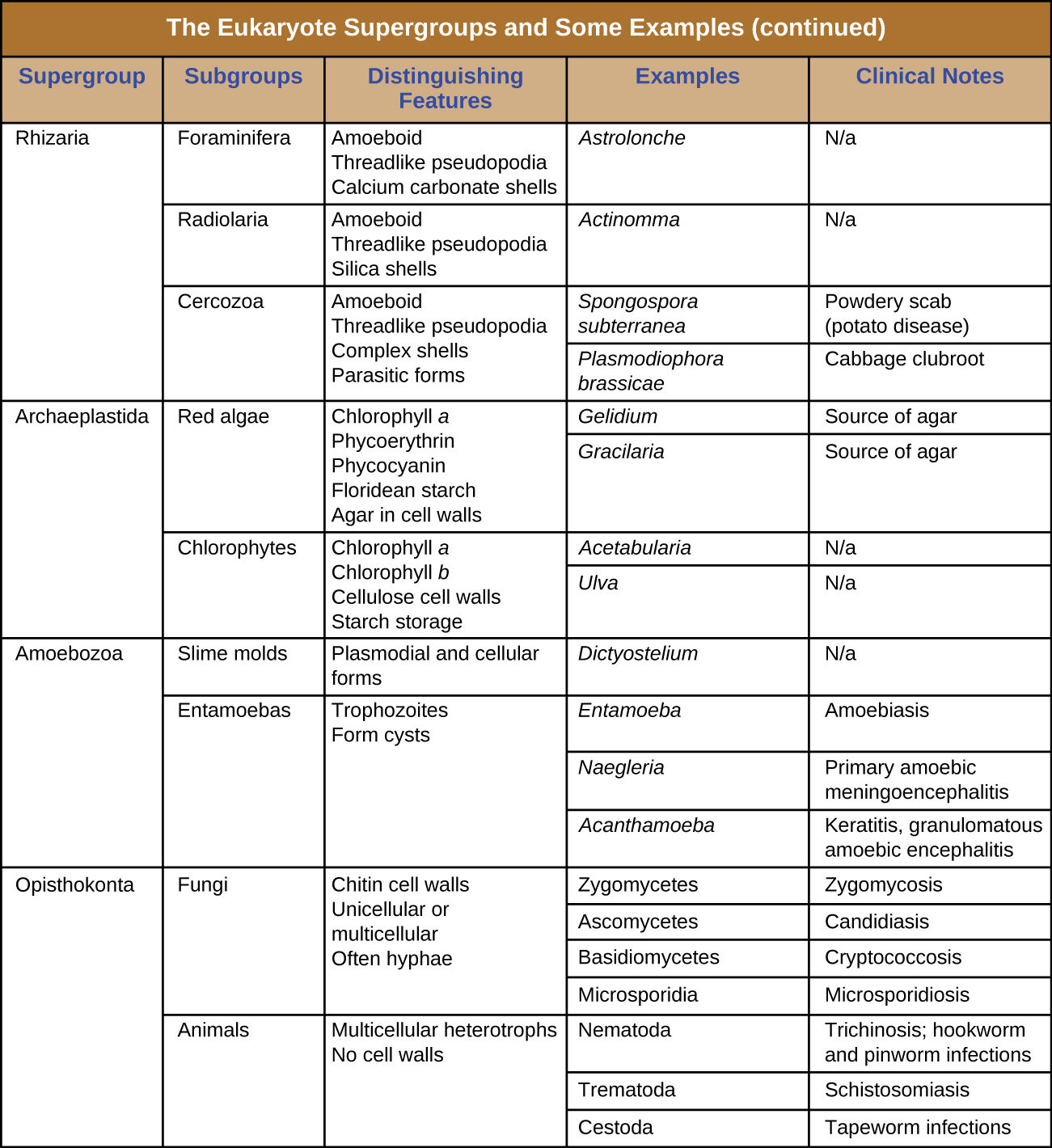
The protists are a polyphyletic group, meaning they lack a shared evolutionary origin. Since the current taxonomy is based on evolutionary history (as determined by biochemistry, morphology, and genetics), protists are scattered across many different taxonomic groups within the domain Eukarya. Eukarya is currently divided into six supergroups that are further divided into subgroups, as illustrated in ( [link] ). In this section, we will primarily be concerned with the supergroups Amoebozoa , Excavata , and Chromalveolata ; these supergroups include many protozoans of clinical significance. The supergroups Opisthokonta and Rhizaria also include some protozoans, but few of clinical significance. In addition to protozoans, Opisthokonta also includes animals and fungi, some of which we will discuss in Parasitic Helminths and Fungi . Some examples of the Archaeplastida will be discussed in Algae . [link] and [link] summarize the characteristics of each supergroup and subgroup and list representatives of each.

A branching tree diagram with common eukaryotic ancestor at the base. This leads to 5 branches. The top branch branches are classified as Excavata which is divided into 3 groups: diplomonads, parabasalids, and euglenozoans. The next branch splits into 2 branches: alveolates, and stramenopiles. The alveolates are divided into dinoflagellates, apicomplexans and ciliates. The stramenopiles are divided into diatoms, golden algae, brown algae and oomyces. All the alveotate and stramenopile groups are labeled Chromalveolata. The next branch divides into cercozoans, forams and radiolarians. These are all labeled rhizaria. The next branch divides into the red algae, chlorophytes (green algae), charophytes (green algae) and land plant. Thesea re all labeldd archaeplastidia. The next branch splits into 2. The top branch divides into slime molds, gymnamoebas and entamoebas. These are all labeled amoebozoa. The bottom branch divides into nucleariids, fungi, choanoflagellates, and animals. These are all labeled  opisthokonta.
This tree shows a proposed classification of the domain Eukarya based on evolutionary relationships. Currently, the domain Eukarya is divided into six supergroups. Within each supergroup are multiple kingdoms. Dotted lines indicate suggested evolutionary relationships that remain under debate.
Table titled: the eukaryote supergroups and some example species. There are 5 columns in the table: supergroup, subgroup, distinguishing features, examples and clinical notes. Supergroup Exacavata is divided into 3 subgroups: fornicate, parabasalids, euglenozoans. Fornicata have the following distinguishing features: form cysts, pair of equal nuclei, no mitochondria, often parasitic, four free flagella. An example is giardia lamblia which causes giardiasis. Parabasalids have the following distinguishing features: no mitochondria, four free flagella, one attached flagellum, no cysts, parasitic or symbiotic, basal bodies, kinetoplastids. An example is Trichomonas which causes trichomoniasis. Euglenozoans have the following distinguishing features: photosynthetic or heterotrophic, flagella. Examples include: Euglena which does not cause disease, Trypanosoma which causes African sleeping sickness and Chagas disease, Leishmanial which causes leishmaniasis. The supergroup Chromalveolata is divided into 4 subgroups: dinoflagellates, apicomplexans, ciliates, and oomyctes/peronosporomycetes. Dinoflagellates have the following distinguishing features: cellulose theca and two dissimilar flagella. Examples include Gonyaulax which causes red tides, Alexandrium which causes paralytic shellfish poisoning, and Pfiesteria which causes harmful algal blooms. Apicomplexans have the following distinguishing features: intracellular parasite and apical organelles. Examples include Plasmodium which causes malaria, Cryptosporidium which causes cryptosporidiosis, Theileria (Babesia) which causes babeiosis, and Toxoplasma which causes Tosoplasmosis. Ciliates have the characteristic of cilia. Examples include Balantidium which causes Balantidiasis. Paramecium and Stentor which do not cause diseas. Oomycetes / peronosporomycetes have the following distinguishing features: water molds, generally diploid, cellulose cell wall. An example is Phytophthora which causes diseases in crops.
Table titled: the eukaryote supergroups and some example species. There are 5 columns in the table: supergroup, subgroup, distinguishing features, examples and clinical notes. The supergroup Rhizaria is divided into 3 subroups: Foraminifera, Radiolaria, and Cerozoa. Foraminifer have the following distinguishing features: amoeboid, thereadlike pseudopodia, calclium carbonate shells. An example is Astrolonche which does not cause disease. Radiolaria have the following distinguishing features: amoeboid, threadlike pseudopodia, silica shells. An example is Actinomma which does not cause disease. Cerozoa have the following distinguishing features: amoeboid, threadlike pseudopodia, complex shells, parasitic forms. Examples include Spongospora subterranean which causes powdery scab (potato disease) and Plasmodiophora brassicae which causes cabbage clubroot. Supergroup Archaeplastida is divided into 2 groups: red algae and Chlorophytes. Red algae have the following distinguishing features: chlorophyll a, phycoerythrin, phycocyanin, flodean starch, agar in cell walls. Examples include Gelidium and Gracilaria which are sources of agar. Chlorophytes have the following distinguishing features: chlorphyll a, chlorophyll b, cellulose cell walls, starch storage. Examples include Acetabularia and Ulva which do not cause disease. Supergroup Amoebozoa is divided into 2 subgroups: slime molds and entamoebas. Slime molds have plasmodial and cellular forms. An example is Dictyostelium which does not cause disease. Entamoebas have the following distinguishing features: trophozoites and form cysts. Examples include Entamoeba which causes Amoebiasis, Naegleria which causes Primary amoebic meningoencephalitis, and Acanthamoeba which causes Keratitis, and granulomatous ameoebic encephalitis. Supergroup Opisthokonta is divided into subroups fungin and animals. Fungi have the following distinguishing features: chitin cell walls, unicellular or multicellular, often hyphae. Examples include Zygomyctes which cause zygomycosis, Asomycetes which cause Candidiasis, Basidiomycetes which cause Cryptococcosis, and Microsporidia which causes microsporidiosis. Animals have the following distinguishing features: multicellular heterotrophs with no cell walls. Examples include Nematoda which cause Trichonosis, hookworm and pinworm infections, Termatoda which causes Schistosomiais, and Cestoda which causes tapeworm infection.
  • Which supergroups contain the clinically significant protists?

Amoebozoa

The supergroup Amoebozoa includes protozoans that use amoeboid movement. Actin microfilaments produce pseudopodia , into which the remainder of the protoplasm flows, thereby moving the organism. The genus Entamoeba includes commensal or parasitic species, including the medically important E. histolytica , which is transmitted by cysts in feces and is the primary cause of amoebic dysentery . The notorious “brain-eating amoeba,” Naegleria fowleri , is also classified within the Amoebozoa. This deadly parasite is found in warm, fresh water and causes primary amoebic meningoencephalitis (PAM) . Another member of this group is Acanthamoeba , which can cause keratitis (corneal inflammation) and blindness.

Questions & Answers

prostaglandin and fever
Maha Reply
Discuss the differences between taste and flavor, including how other sensory inputs contribute to our  perception of flavor.
John Reply
taste refers to your understanding of the flavor . while flavor one The other hand is refers to sort of just a blend things.
Faith
While taste primarily relies on our taste buds, flavor involves a complex interplay between taste and aroma
Kamara
which drugs can we use for ulcers
Ummi Reply
omeprazole
Kamara
what
Renee
what is this
Renee
is a drug
Kamara
of anti-ulcer
Kamara
Omeprazole Cimetidine / Tagament For the complicated once ulcer - kit
Patrick
what is the function of lymphatic system
Nency Reply
Not really sure
Eli
to drain extracellular fluid all over the body.
asegid
The lymphatic system plays several crucial roles in the human body, functioning as a key component of the immune system and contributing to the maintenance of fluid balance. Its main functions include: 1. Immune Response: The lymphatic system produces and transports lymphocytes, which are a type of
asegid
to transport fluids fats proteins and lymphocytes to the blood stream as lymph
Adama
what is anatomy
Oyindarmola Reply
Anatomy is the identification and description of the structures of living things
Kamara
what's the difference between anatomy and physiology
Oyerinde Reply
Anatomy is the study of the structure of the body, while physiology is the study of the function of the body. Anatomy looks at the body's organs and systems, while physiology looks at how those organs and systems work together to keep the body functioning.
AI-Robot
what is enzymes all about?
Mohammed Reply
Enzymes are proteins that help speed up chemical reactions in our bodies. Enzymes are essential for digestion, liver function and much more. Too much or too little of a certain enzyme can cause health problems
Kamara
yes
Prince
how does the stomach protect itself from the damaging effects of HCl
Wulku Reply
little girl okay how does the stomach protect itself from the damaging effect of HCL
Wulku
it is because of the enzyme that the stomach produce that help the stomach from the damaging effect of HCL
Kamara
function of digestive system
Ali Reply
function of digestive
Ali
the diagram of the lungs
Adaeze Reply
what is the normal body temperature
Diya Reply
37 degrees selcius
Xolo
37°c
Stephanie
please why 37 degree selcius normal temperature
Mark
36.5
Simon
37°c
Iyogho
the normal temperature is 37°c or 98.6 °Fahrenheit is important for maintaining the homeostasis in the body the body regular this temperature through the process called thermoregulation which involves brain skin muscle and other organ working together to maintain stable internal temperature
Stephanie
37A c
Wulku
what is anaemia
Diya Reply
anaemia is the decrease in RBC count hemoglobin count and PVC count
Eniola
what is the pH of the vagina
Diya Reply
how does Lysin attack pathogens
Diya
acid
Mary
I information on anatomy position and digestive system and there enzyme
Elisha Reply
anatomy of the female external genitalia
Muhammad Reply
Got questions? Join the online conversation and get instant answers!
Jobilize.com Reply

Get Jobilize Job Search Mobile App in your pocket Now!

Get it on Google Play Download on the App Store Now


Source:  OpenStax, Microbiology. OpenStax CNX. Nov 01, 2016 Download for free at http://cnx.org/content/col12087/1.4
Google Play and the Google Play logo are trademarks of Google Inc.

Notification Switch

Would you like to follow the 'Microbiology' conversation and receive update notifications?

Ask