<< Chapter < Page Chapter >> Page >

Learning objectives

  • Describe the unique features of nonproteobacteria gram-negative bacteria
  • Give an example of a nonproteobacteria bacterium in each category
  • Describe the unique features of phototrophic bacteria
  • Identify phototrophic bacteria

The majority of the gram-negative bacteria belong to the phylum Proteobacteria, discussed in the previous section. Those that do not are called the nonproteobacteria . In this section, we will describe three classes of gram-negative nonproteobacteria: the spirochetes, the CFB group , and the Planctomycetes . A diverse group of phototrophic bacteria that includes Proteobacteria and nonproteobacteria will be discussed at the end of this section.

Spirochetes

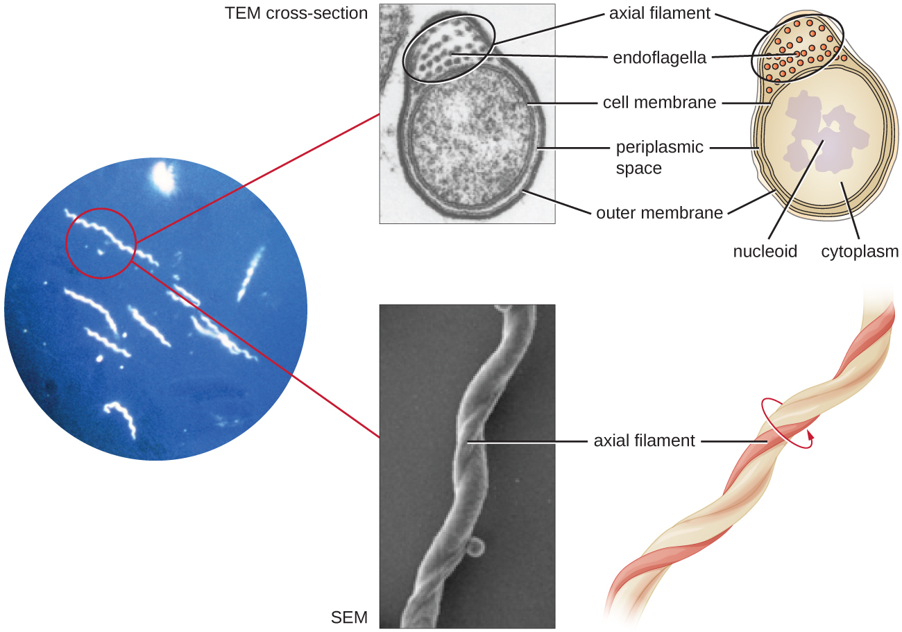
Spirochetes are characterized by their long (up to 250 μm), spiral-shaped bodies. Most spirochetes are also very thin, which makes it difficult to examine gram-stained preparations under a conventional brightfield microscope. Darkfield fluorescent microscopy is typically used instead. Spirochetes are also difficult or even impossible to culture. They are highly motile, using their axial filament to propel themselves. The axial filament is similar to a flagellum, but it wraps around the cell and runs inside the cell body of a spirochete in the periplasmic space between the outer membrane and the plasma membrane ( [link] ).

A light micrograph of long spiral shaped cells. A TEM cross-section of these shows a circle outlined by a cell membrane. Inside the cell is the cytoplasm and a darker region labeled nucleoid. Outside of this is the periplasmic space and outside of that is an outer membrane. A bulge within the periplasmic space is labeled axial filament. Small dots within the axial filament are labeled endoflagella. An SEM from the original light micrograph shows what looks like a thin rope wound around a thicker rope. The thin rope is labeled axial filament.
Spirochetes are typically observed using darkfield microscopy (left). However, electron microscopy (top center, bottom center) provides a more detailed view of their cellular morphology. The flagella found between the inner and outer membranes of spirochetes wrap around the bacterium, causing a twisting motion used for locomotion. (credit “spirochetes” micrograph: modification of work by Centers for Disease Control and Prevention; credit “SEM/TEM”: modification of work by Guyard C, Raffel SJ, Schrumpf ME, Dahlstrom E, Sturdevant D, Ricklefs SM, Martens C, Hayes SF, Fischer ER, Hansen BT, Porcella SF, Schwan TG)

Several genera of spirochetes include human pathogens. For example, the genus Treponema includes a species T. pallidum , which is further classified into four subspecies: T. pallidum pallidum , T. pallidum pertenue, T. pallidum carateum, and T. pallidum endemicum . The subspecies T. pallidum pallidum causes the sexually transmitted infection known as syphilis , the third most prevalent sexually transmitted bacterial infection in the United States, after chlamydia and gonorrhea. The other subspecies of T. pallidum cause tropical infectious diseases of the skin, bones, and joints.

Another genus of spirochete, Borrelia , contains a number of pathogenic species. B. burgdorferi causes Lyme disease , which is transmitted by several genera of ticks (notably Ixodes and Amblyomma ) and often produces a “bull’s eye” rash, fever, fatigue, and, sometimes, debilitating arthritis. B. recurrens causes a condition known as relapsing fever. Appendix D lists the genera, species, and related diseases for spirochetes.

  • Why do scientists typically use darkfield fluorescent microscopy to visualize spirochetes?

Cytophaga , Fusobacterium , and Bacteroides

The gram-negative nonproteobacteria of the genera Cytophaga , Fusobacterium , and Bacteroides are classified together as a phylum and called the CFB group . Although they are phylogenetically diverse, bacteria of the CFB group share some similarities in the sequence of nucleotides in their DNA. They are rod-shaped bacteria adapted to anaerobic environments, such as the tissue of the gums, gut, and rumen of ruminating animals. CFB bacteria are avid fermenters, able to process cellulose in rumen, thus enabling ruminant animals to obtain carbon and energy from grazing.

Questions & Answers

what is biology
Hajah Reply
the study of living organisms and their interactions with one another and their environments
AI-Robot
what is biology
Victoria Reply
HOW CAN MAN ORGAN FUNCTION
Alfred Reply
the diagram of the digestive system
Assiatu Reply
allimentary cannel
Ogenrwot
How does twins formed
William Reply
They formed in two ways first when one sperm and one egg are splited by mitosis or two sperm and two eggs join together
Oluwatobi
what is genetics
Josephine Reply
Genetics is the study of heredity
Misack
how does twins formed?
Misack
What is manual
Hassan Reply
discuss biological phenomenon and provide pieces of evidence to show that it was responsible for the formation of eukaryotic organelles
Joseph Reply
what is biology
Yousuf Reply
the study of living organisms and their interactions with one another and their environment.
Wine
discuss the biological phenomenon and provide pieces of evidence to show that it was responsible for the formation of eukaryotic organelles in an essay form
Joseph Reply
what is the blood cells
Shaker Reply
list any five characteristics of the blood cells
Shaker
lack electricity and its more savely than electronic microscope because its naturally by using of light
Abdullahi Reply
advantage of electronic microscope is easily and clearly while disadvantage is dangerous because its electronic. advantage of light microscope is savely and naturally by sun while disadvantage is not easily,means its not sharp and not clear
Abdullahi
cell theory state that every organisms composed of one or more cell,cell is the basic unit of life
Abdullahi
is like gone fail us
DENG
cells is the basic structure and functions of all living things
Ramadan
What is classification
ISCONT Reply
is organisms that are similar into groups called tara
Yamosa
in what situation (s) would be the use of a scanning electron microscope be ideal and why?
Kenna Reply
A scanning electron microscope (SEM) is ideal for situations requiring high-resolution imaging of surfaces. It is commonly used in materials science, biology, and geology to examine the topography and composition of samples at a nanoscale level. SEM is particularly useful for studying fine details,
Hilary
cell is the building block of life.
Condoleezza Reply
what is phylogeny
Odigie Reply
evolutionary history and relationship of an organism or group of organisms
AI-Robot
Got questions? Join the online conversation and get instant answers!
Jobilize.com Reply
Practice MCQ 3

Get Jobilize Job Search Mobile App in your pocket Now!

Get it on Google Play Download on the App Store Now


Source:  OpenStax, Microbiology. OpenStax CNX. Nov 01, 2016 Download for free at http://cnx.org/content/col12087/1.4
Google Play and the Google Play logo are trademarks of Google Inc.

Notification Switch

Would you like to follow the 'Microbiology' conversation and receive update notifications?

Ask