<< Chapter < Page Chapter >> Page >

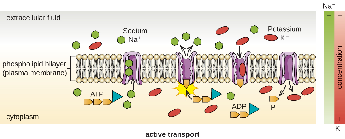
Active transport occurs when cells move molecules across their membrane against concentration gradients ( [link] ). A major difference between passive and active transport is that active transport requires adenosine triphosphate (ATP) or other forms of energy to move molecules “uphill.” Therefore, active transport structures are often called “pumps.”
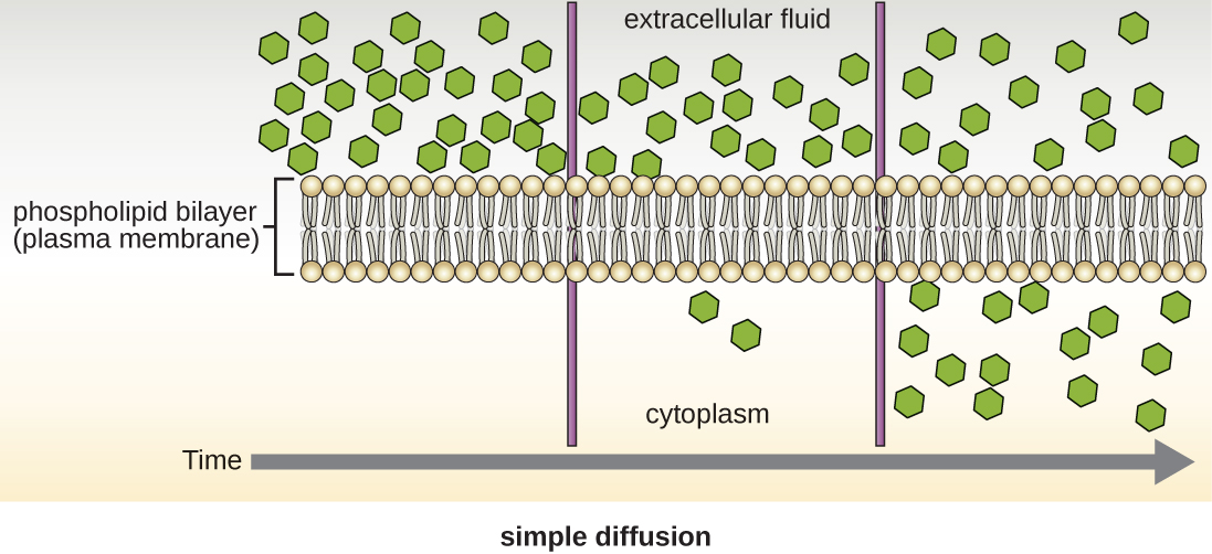
Simple diffusion. A diagram with a phospholipid bilayer (plasma membrane) along the middle. Above the bilayer is the extracellular fluid and below is the cytoplasm. At the far left there are many hexagons in the extracellular fluid above the bilayer and none in the cytoplasm below. At a later time shown in the middle of the timeline there are a few hexagons in the cytoplasm and still many in the extracellular fluid. At the last timeframe shown on the right there are equal numbers of hexagons in the extracellular fluid as in the cytoplasm.
Simple diffusion down a concentration gradient directly across the phospholipid bilayer. (credit: modification of work by Mariana Ruiz Villareal)
Facilitated diffusion. A diagram with a phospholipid bilayer (plasma membrane) in the middle of the image. There are many hexagons in the extracellular fluid above the membrane and few hexagons in the cytoplasm below the membrane. A protein channel is shown transporting the hexagons across the membrane from the extracellular fluid to the cytoplasm.
Facilitated diffusion down a concentration gradient through a membrane protein. (credit: modification of work by Mariana Ruiz Villareal)
Active Transport. A diagram with a phospholipid bilayer (plasma membrane) along the middle. Above the bilayer is the extracellular fluid and below is the cytoplasm. There are more sodium ions in the extracellular fluid than in the cytoplasm. There are more potassium ions in the cytoplasm than in the extracellular fluid. A protein in the membrane is shown moving sodium from the cytoplasm to the extracellular fluid. The same membrane is shown moving potassium from the extracellular fluid to the cytoplasm. As the protein moves these ions, it also breaks down ATP to ADP.
Active transport against a concentration gradient via a membrane pump that requires energy. (credit: modification of work by Mariana Ruiz Villareal)

Group translocation also transports substances into bacterial cells. In this case, as a molecule moves into a cell against its concentration gradient, it is chemically modified so that it does not require transport against an unfavorable concentration gradient. A common example of this is the bacterial phosphotransferase system, a series of carriers that phosphorylates (i.e., adds phosphate ions to) glucose or other sugars upon entry into cells. Since the phosphorylation of sugars is required during the early stages of sugar metabolism, the phosphotransferase system is considered to be an energy neutral system.

Photosynthetic membrane structures

Some prokaryotic cells, namely cyanobacteria and photosynthetic bacteria , have membrane structures that enable them to perform photosynthesis. These structures consist of an infolding of the plasma membrane that encloses photosynthetic pigments such as green chlorophylls and bacteriochlorophylls . In cyanobacteria, these membrane structures are called thylakoids; in photosynthetic bacteria, they are called chromatophores, lamellae, or chlorosomes.

Cell wall

The primary function of the cell wall is to protect the cell from harsh conditions in the outside environment. When present, there are notable similarities and differences among the cell walls of archaea, bacteria, and eukaryotes.

The major component of bacterial cell walls is called peptidoglycan (or murein ); it is only found in bacteria. Structurally, peptidoglycan resembles a layer of meshwork or fabric ( [link] ). Each layer is composed of long chains of alternating molecules of N-acetylglucosamine (NAG) and N-acetylmuramic acid (NAM). The structure of the long chains has significant two-dimensional tensile strength due to the formation of peptide bridges that connect NAG and NAM within each peptidoglycan layer. In gram-negative bacteria, tetrapeptide chains extending from each NAM unit are directly cross-linked, whereas in gram-positive bacteria, these tetrapeptide chains are linked by pentaglycine cross-bridges. Peptidoglycan subunits are made inside of the bacterial cell and then exported and assembled in layers, giving the cell its shape.

Since peptidoglycan is unique to bacteria, many antibiotic drugs are designed to interfere with peptidoglycan synthesis, weakening the cell wall and making bacterial cells more susceptible to the effects of osmotic pressure (see Mechanisms of Antibacterial Drugs ). In addition, certain cells of the human immune system are able “recognize” bacterial pathogens by detecting peptidoglycan on the surface of a bacterial cell; these cells then engulf and destroy the bacterial cell, using enzymes such as lysozyme, which breaks down and digests the peptidoglycan in their cell walls (see Pathogen Recognition and Phagocytosis ).

Questions & Answers

what is biology
Hajah Reply
the study of living organisms and their interactions with one another and their environments
AI-Robot
what is biology
Victoria Reply
HOW CAN MAN ORGAN FUNCTION
Alfred Reply
the diagram of the digestive system
Assiatu Reply
allimentary cannel
Ogenrwot
How does twins formed
William Reply
They formed in two ways first when one sperm and one egg are splited by mitosis or two sperm and two eggs join together
Oluwatobi
what is genetics
Josephine Reply
Genetics is the study of heredity
Misack
how does twins formed?
Misack
What is manual
Hassan Reply
discuss biological phenomenon and provide pieces of evidence to show that it was responsible for the formation of eukaryotic organelles
Joseph Reply
what is biology
Yousuf Reply
the study of living organisms and their interactions with one another and their environment.
Wine
discuss the biological phenomenon and provide pieces of evidence to show that it was responsible for the formation of eukaryotic organelles in an essay form
Joseph Reply
what is the blood cells
Shaker Reply
list any five characteristics of the blood cells
Shaker
lack electricity and its more savely than electronic microscope because its naturally by using of light
Abdullahi Reply
advantage of electronic microscope is easily and clearly while disadvantage is dangerous because its electronic. advantage of light microscope is savely and naturally by sun while disadvantage is not easily,means its not sharp and not clear
Abdullahi
cell theory state that every organisms composed of one or more cell,cell is the basic unit of life
Abdullahi
is like gone fail us
DENG
cells is the basic structure and functions of all living things
Ramadan
What is classification
ISCONT Reply
is organisms that are similar into groups called tara
Yamosa
in what situation (s) would be the use of a scanning electron microscope be ideal and why?
Kenna Reply
A scanning electron microscope (SEM) is ideal for situations requiring high-resolution imaging of surfaces. It is commonly used in materials science, biology, and geology to examine the topography and composition of samples at a nanoscale level. SEM is particularly useful for studying fine details,
Hilary
cell is the building block of life.
Condoleezza Reply
Got questions? Join the online conversation and get instant answers!
Jobilize.com Reply

Get Jobilize Job Search Mobile App in your pocket Now!

Get it on Google Play Download on the App Store Now


Source:  OpenStax, Microbiology. OpenStax CNX. Nov 01, 2016 Download for free at http://cnx.org/content/col12087/1.4
Google Play and the Google Play logo are trademarks of Google Inc.

Notification Switch

Would you like to follow the 'Microbiology' conversation and receive update notifications?

Ask