<< Chapter < Page Chapter >> Page >
a) Diagram of the circulatory system. Blood from the lower part of the body travels to the heart in the inferior vena cava. Blood from the upper part of the body travels to the heart in the superior vena cava. The pulmonary artery travels from the heart to the lungs and the pulmonary vein travels from the lungs to the heart. B) A diagram of the heart showing the flow of blood. Beginning in the superior and inferior vena cavas, blood travels to the right atrium, through the tricuspid valve, to the right ventricle and out the pulmonary artery. The image does not follow the pulmonary artery. From the pulmonary vein blood travels to the left atrium, through the mitral valve, to the left ventricle and out the aorta.  C) The layers of the heart. The Pericardium is the outer layer. The myocardium is the thick middle layer. The endocardium is the inner layer.
The major components of the human circulatory system include the heart, arteries, veins, and capillaries. This network delivers blood to the body’s organs and tissues. (credit top left: modification of work by Mariana Ruiz Villareal; credit bottom right: modification of work by Bruce Blaus)

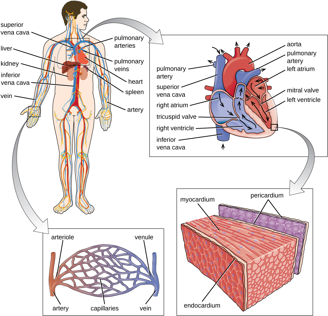
Other organs play important roles in the circulatory system as well. The kidneys filter the blood, removing waste products and eliminating them in the urine. The liver also filters the blood and removes damaged or defective red blood cells. The spleen filters and stores blood, removes damaged red blood cells, and is a reservoir for immune factors. All of these filtering structures serve as sites for entrapment of microorganisms and help maintain an environment free of microorganisms in the blood.

The lymphatic system

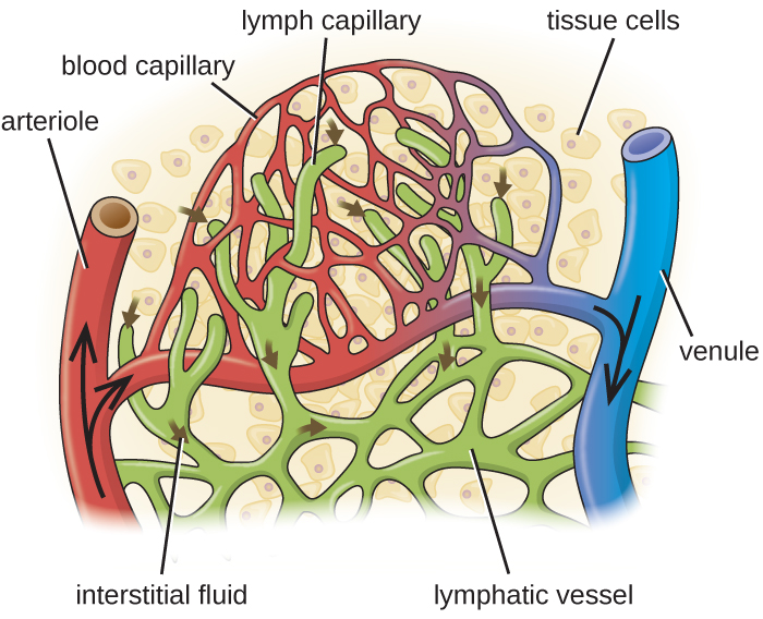
The lymphatic system is also a network of vessels that run throughout the body ( [link] ). However, these vessels do not form a full circulating system and are not pressurized by the heart. Rather, the lymphatic system is an open system with the fluid moving in one direction from the extremities toward two drainage points into veins just above the heart. Lymphatic fluids move more slowly than blood because they are not pressurized. Small lymph capillaries interact with blood capillaries in the interstitial spaces in tissues. Fluids from the tissues enter the lymph capillaries and are drained away ( [link] ). These fluids, termed lymph , also contain large numbers of white blood cells.

Diagram of the lymphatic system. Lymph notes are swellings on tubes (called lymph vessels) that travel throughout the body. The right lymphatic duct and entering vein are in the neck. A tonsil is a swelling on the lymph vessel in the mouth. The thymus is a lumpy structure on the heart. A close-up of a lymph node shows a roundish structure with many tubes attached to it. The central area has a box labeled “masses of lymphocytes and macrophages”. A close-up of this area shows tissue cells in the background with a blood capillary network. Lymph vessels run between the  blood capillary network. Lymphatic capillaries are the ends of the lymph vessels. Fluid from around the cells (called interstitial fluid) enters the lymphatic capillaries and travels through the lymphatic vessels.
The essential components of the human lymphatic system drain fluid away from tissues.
Blood enters the capillaries from an arteriole (red) and leaves through venules (blue). Interstitial fluids may drain into the lymph capillaries (green) and proceed to lymph nodes. A close-up of tissue cells in interstitial fluid. An arteriole and a venule are connected by a network of capillaries. Lymphatic vessels are also a network  in this region and end in lymph capillarie.
Blood enters the capillaries from an arteriole (red) and leaves through venules (blue). Interstitial fluids may drain into the lymph capillaries (green) and proceed to lymph nodes. (credit: modification of work by National Cancer Institute, National Institutes of Health)

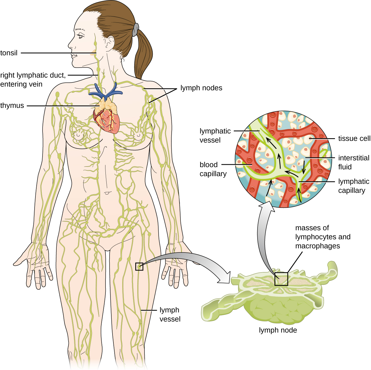
The lymphatic system contains two types of lymphoid tissues. The primary lymphoid tissue includes bone marrow and the thymus. Bone marrow contains the hematopoietic stem cells (HSC) that differentiate and mature into the various types of blood cells and lymphocytes (see [link] ). The secondary lymphoid tissue s include the spleen, lymph nodes, and several areas of diffuse lymphoid tissues underlying epithelial membranes. The spleen , an encapsulated structure, filters blood and captures pathogens and antigens that pass into it ( [link] ). The spleen contains specialized macrophages and dendritic cells that are crucial for antigen presentation, a mechanism critical for activation of T lymphocytes and B lymphocytes (see Major Histocompatibility Complexes and Antigen-Presenting Cells ). Lymph nodes are bean-shaped organs situated throughout the body. These structures contain areas called germinal centers that are rich in B and T lymphocytes. The lymph nodes also contain macrophages and dendritic cells for antigen presentation. Lymph from nearby tissues enters the lymph node through afferent lymphatic vessels and encounters these lymphocytes as it passes through; the lymph exits the lymph node through the efferent lymphatic vessels ( [link] ).

Questions & Answers

what is biology
Hajah Reply
the study of living organisms and their interactions with one another and their environments
AI-Robot
what is biology
Victoria Reply
HOW CAN MAN ORGAN FUNCTION
Alfred Reply
the diagram of the digestive system
Assiatu Reply
allimentary cannel
Ogenrwot
How does twins formed
William Reply
They formed in two ways first when one sperm and one egg are splited by mitosis or two sperm and two eggs join together
Oluwatobi
what is genetics
Josephine Reply
Genetics is the study of heredity
Misack
how does twins formed?
Misack
What is manual
Hassan Reply
discuss biological phenomenon and provide pieces of evidence to show that it was responsible for the formation of eukaryotic organelles
Joseph Reply
what is biology
Yousuf Reply
the study of living organisms and their interactions with one another and their environment.
Wine
discuss the biological phenomenon and provide pieces of evidence to show that it was responsible for the formation of eukaryotic organelles in an essay form
Joseph Reply
what is the blood cells
Shaker Reply
list any five characteristics of the blood cells
Shaker
lack electricity and its more savely than electronic microscope because its naturally by using of light
Abdullahi Reply
advantage of electronic microscope is easily and clearly while disadvantage is dangerous because its electronic. advantage of light microscope is savely and naturally by sun while disadvantage is not easily,means its not sharp and not clear
Abdullahi
cell theory state that every organisms composed of one or more cell,cell is the basic unit of life
Abdullahi
is like gone fail us
DENG
cells is the basic structure and functions of all living things
Ramadan
What is classification
ISCONT Reply
is organisms that are similar into groups called tara
Yamosa
in what situation (s) would be the use of a scanning electron microscope be ideal and why?
Kenna Reply
A scanning electron microscope (SEM) is ideal for situations requiring high-resolution imaging of surfaces. It is commonly used in materials science, biology, and geology to examine the topography and composition of samples at a nanoscale level. SEM is particularly useful for studying fine details,
Hilary
cell is the building block of life.
Condoleezza Reply
Got questions? Join the online conversation and get instant answers!
Jobilize.com Reply

Get Jobilize Job Search Mobile App in your pocket Now!

Get it on Google Play Download on the App Store Now


Source:  OpenStax, Microbiology. OpenStax CNX. Nov 01, 2016 Download for free at http://cnx.org/content/col12087/1.4
Google Play and the Google Play logo are trademarks of Google Inc.

Notification Switch

Would you like to follow the 'Microbiology' conversation and receive update notifications?

Ask