

| << Chapter < Page | Chapter >> Page > |
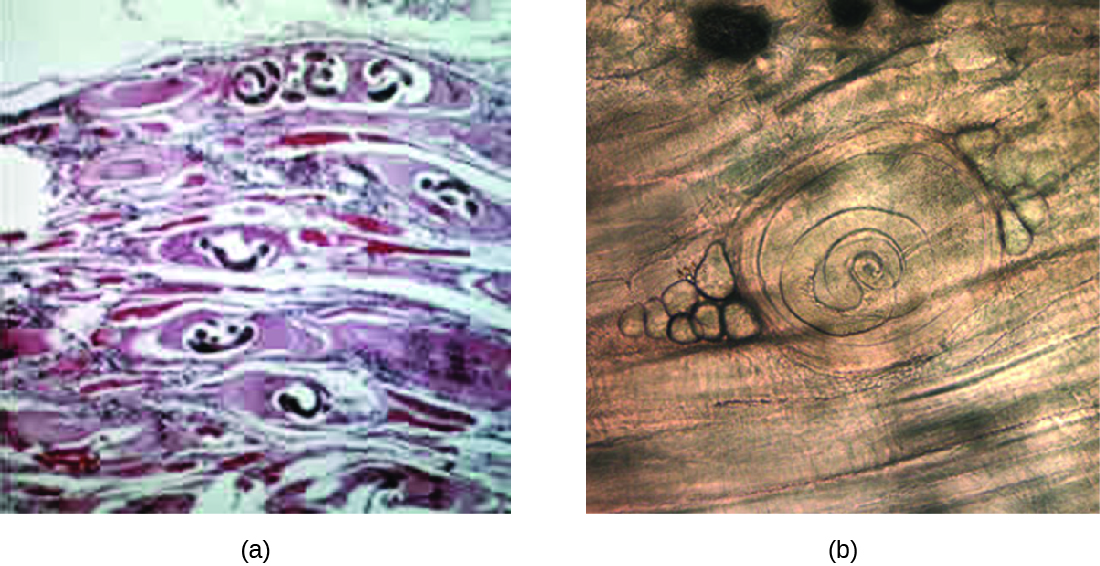
Infection is diagnosed using clinical history, muscle biopsy to look for larvae, and serological testing, including immunoassays. Enzyme immunoassay is the most common test. It is difficult to effectively treat larvae that have formed cysts in the muscle, although medications may help. It is best to begin treatment as soon as possible because medications such as mebendazole and albendazole are effective in killing only the adult worms in the intestine. Steroids may be used to reduce inflammation if larvae are in the muscles.
Taeniasis is a tapeworm infection, generally caused by pork ( Taenia solium ), beef ( Taenia saginata ), and Asian ( Taenia asiatica ) tapeworms found in undercooked meat. Consumption of raw or undercooked fish, including contaminated sushi, can also result in infection from the fish tapeworm ( Diphyllobothrium latum ). Tapeworms are flatworms ( cestodes ) with multiple body segments and a head called a scolex that attaches to the intestinal wall. Tapeworms can become quite large, reaching 4 to 8 meters long ( [link] ). [link] illustrates the life cycle of a tapeworm.
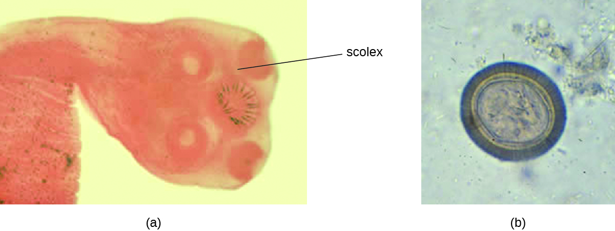
Tapeworms attached to the intestinal wall produce eggs that are excreted in feces. After ingestion by animals, the eggs hatch and the larvae emerge. They may take up residence in the intestine, but can sometimes move to other tissues, especially muscle or brain tissue. When T. solium larvae form cysts in tissue, the condition is called cysticercosis . This occurs through ingestion of eggs via the fecal-oral route, not through consumption of undercooked meat. It can develop in the muscles, eye (ophthalmic cysticercosis), or brain (neurocysticercosis).
Infections may be asymptomatic or they may cause mild gastrointestinal symptoms such as epigastric discomfort, nausea, diarrhea, flatulence, or hunger pains. It is also common to find visible tapeworm segments passed in the stool. In cases of cysticercosis, symptoms differ depending upon where the cysts become established. Neurocysticercosis can have severe, life-threatening consequences and is associated with headaches and seizures because of the presence of the tapeworm larvae encysted in the brain. Cysts in muscles may be asymptomatic, or they may be painful.
To diagnose these conditions, microscopic analysis of stool samples from three separate days is generally recommended. Eggs or body segments, called proglottids, may be visible in these samples. Molecular methods have been developed but are not yet widely available. Imaging, such as CT and MRI, may be used to detect cysts. Praziquantel or niclosamide are used for treatment.
Notification Switch
Would you like to follow the 'Microbiology' conversation and receive update notifications?

|
|

|
|

|
|

|
|

|
|

|
|

|
|

|
|

|
|

|