<< Chapter < Page Chapter >> Page >
Structure of a tooth. The part above the gums (gingiva) is called the crown, the part below the gingiva is the root. The very top of the crown is a thick enamel, this is very thick at the region above the gingiva and much thinner in the root. The next layer in is the dentin and this is equally thick in both the crown and root. In the very center is the pulp which contains the pulp canal (root canal) and nerve and blood vessels. The root sits mainly in the bone region. There is a small space where the tooth extends past the gingiva, this is called the gingival crevice.
The tooth has a visible crown with an outer layer of enamel, a layer of dentin, and an inner pulp. The root, hidden by the gums, contains the pulp canal (root canal). (credit: modification of work by Bruce Blaus)

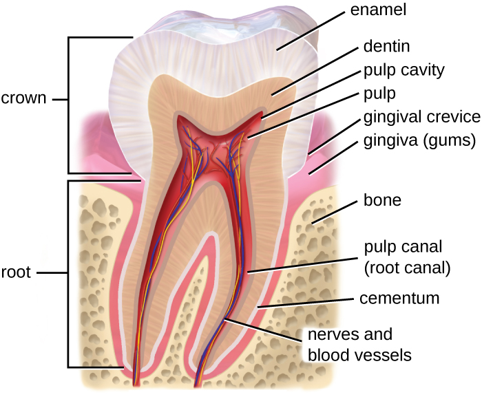
Microbes such as bacteria and archaea are abundant in the mouth and coat all of the surfaces of the oral cavity. However, different structures, such as the teeth or cheeks, host unique communities of both aerobic and anaerobic microbes. Some factors appear to work against making the mouth hospitable to certain microbes. For example, chewing allows microbes to mix better with saliva so they can be swallowed or spit out more easily. Saliva also contains enzymes, including lysozyme , which can damage microbial cells. Recall that lysozyme is part of the first line of defense in the innate immune system and cleaves the β-(1,4) glycosidic linkages between N-acetylglucosamine (NAG) and N-acetylmuramic acid (NAM) in bacterial peptidoglycan (see Chemical Defenses ). Additionally, fluids containing immunoglobulins and phagocytic cells are produced in the gingival spaces. Despite all of these chemical and mechanical activities, the mouth supports a large microbial community.

  • What factors make the mouth inhospitable for certain microbes?

Anatomy and normal microbiota of the gi tract

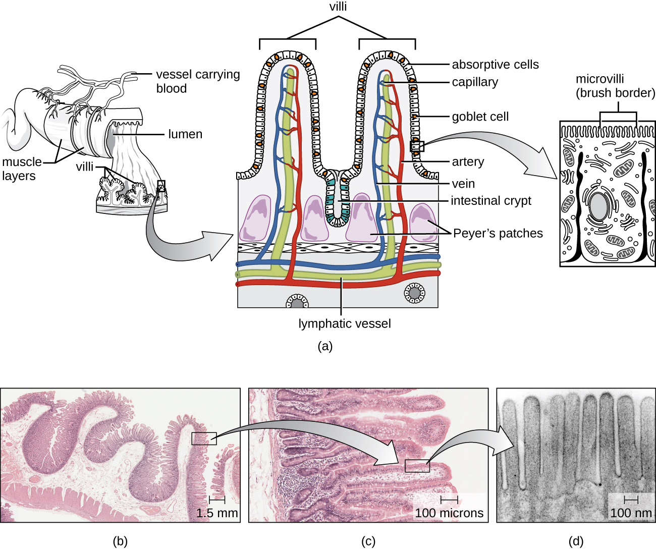
As food leaves the oral cavity, it travels through the pharynx, or the back of the throat, and moves into the esophagus , which carries the food from the pharynx to the stomach without adding any additional digestive enzymes. The stomach produces mucus to protect its lining, as well as digestive enzymes and acid to break down food. Partially digested food then leaves the stomach through the pyloric sphincter , reaching the first part of the small intestine called the duodenum . Pancreatic juice, which includes enzymes and bicarbonate ions, is released into the small intestine to neutralize the acidic material from the stomach and to assist in digestion. Bile, produced by the liver but stored in the gallbladder , is also released into the small intestine to emulsify fats so that they can travel in the watery environment of the small intestine. Digestion continues in the small intestine, where the majority of nutrients contained in the food are absorbed. Simple columnar epithelial cells called enterocytes line the lumen surface of the small intestinal folds called villi . Each enterocyte has smaller microvilli (cytoplasmic membrane extensions) on the cellular apical surface that increase the surface area to allow more absorption of nutrients to occur ( [link] ).

The small intestines with increasing magnification. A) is a diagram and b), c), and d) are micrographs of each magnification. The micrograph of the larges magnification shows a pink region on the bottom with a deeply waved darker pink region at the surface; the top of the image is clear. There are some darker patches in the bottom layer labeled Peyer’s patches. The diagram sows a tube lined with three layers of muscle; blood vessels connected to the outside of the tube. A cutout of the tube shows circular folds along the diameter of the tube. These folds contain deeply lobed villi. The empty space in the tube is labeled lumen. The next layer of magnification is one of the vili. The micrograph is filled with pink layers folding back and forth. The diagram shows two folds. The surface of the fold is covered with absorptive cells and some goblet cells. Between the folds is further indent labeled intestinal crypt. Inside the folds are capillaries, arteries, and lymphatic vesicles. At the very bottom of the structure (below the blood and lymph vessels, are a few duodenal glands. The final close-up shows finger-shapes in a row on the surface of a cell. These are labeled microvilli (brush border) on the diagram.
(a) The structure of the wall of the small intestine allows for the majority of nutrient absorption in the body. (b) Villi are folds in the surface of the small intestine. Microvilli are cytoplasmic extensions on individual cells that increase the surface area for absorption. (c) A light micrograph shows the shape of the villi. (d) An electron micrograph shows the shape of the microvilli. (credit b, c, d: Modification of micrographs provided by the Regents of University of Michigan Medical School © 2012)

Questions & Answers

what is biology
Hajah Reply
the study of living organisms and their interactions with one another and their environments
AI-Robot
what is biology
Victoria Reply
HOW CAN MAN ORGAN FUNCTION
Alfred Reply
the diagram of the digestive system
Assiatu Reply
allimentary cannel
Ogenrwot
How does twins formed
William Reply
They formed in two ways first when one sperm and one egg are splited by mitosis or two sperm and two eggs join together
Oluwatobi
what is genetics
Josephine Reply
Genetics is the study of heredity
Misack
how does twins formed?
Misack
What is manual
Hassan Reply
discuss biological phenomenon and provide pieces of evidence to show that it was responsible for the formation of eukaryotic organelles
Joseph Reply
what is biology
Yousuf Reply
the study of living organisms and their interactions with one another and their environment.
Wine
discuss the biological phenomenon and provide pieces of evidence to show that it was responsible for the formation of eukaryotic organelles in an essay form
Joseph Reply
what is the blood cells
Shaker Reply
list any five characteristics of the blood cells
Shaker
lack electricity and its more savely than electronic microscope because its naturally by using of light
Abdullahi Reply
advantage of electronic microscope is easily and clearly while disadvantage is dangerous because its electronic. advantage of light microscope is savely and naturally by sun while disadvantage is not easily,means its not sharp and not clear
Abdullahi
cell theory state that every organisms composed of one or more cell,cell is the basic unit of life
Abdullahi
is like gone fail us
DENG
cells is the basic structure and functions of all living things
Ramadan
What is classification
ISCONT Reply
is organisms that are similar into groups called tara
Yamosa
in what situation (s) would be the use of a scanning electron microscope be ideal and why?
Kenna Reply
A scanning electron microscope (SEM) is ideal for situations requiring high-resolution imaging of surfaces. It is commonly used in materials science, biology, and geology to examine the topography and composition of samples at a nanoscale level. SEM is particularly useful for studying fine details,
Hilary
cell is the building block of life.
Condoleezza Reply
Got questions? Join the online conversation and get instant answers!
Jobilize.com Reply
Practice MCQ 2

Get Jobilize Job Search Mobile App in your pocket Now!

Get it on Google Play Download on the App Store Now


Source:  OpenStax, Microbiology. OpenStax CNX. Nov 01, 2016 Download for free at http://cnx.org/content/col12087/1.4
Google Play and the Google Play logo are trademarks of Google Inc.

Notification Switch

Would you like to follow the 'Microbiology' conversation and receive update notifications?

Ask