

| << Chapter < Page | Chapter >> Page > |
After a morning of playing outside, four-year-old Carli ran inside for lunch. After taking a bite of her fried egg, she pushed it away and whined, “It’s too slimy, Mommy. I don’t want any more.” But her mother, in no mood for games, curtly replied that if she wanted to go back outside she had better finish her lunch. Reluctantly, Carli complied, trying hard not to gag as she choked down the runny egg.
That night, Carli woke up feeling nauseated. She cried for her parents and then began to vomit. Her parents tried to comfort her, but she continued to vomit all night and began to have diarrhea and run a fever. By the morning, her parents were very worried. They rushed her to the emergency room.
Jump to the next Clinical Focus box.
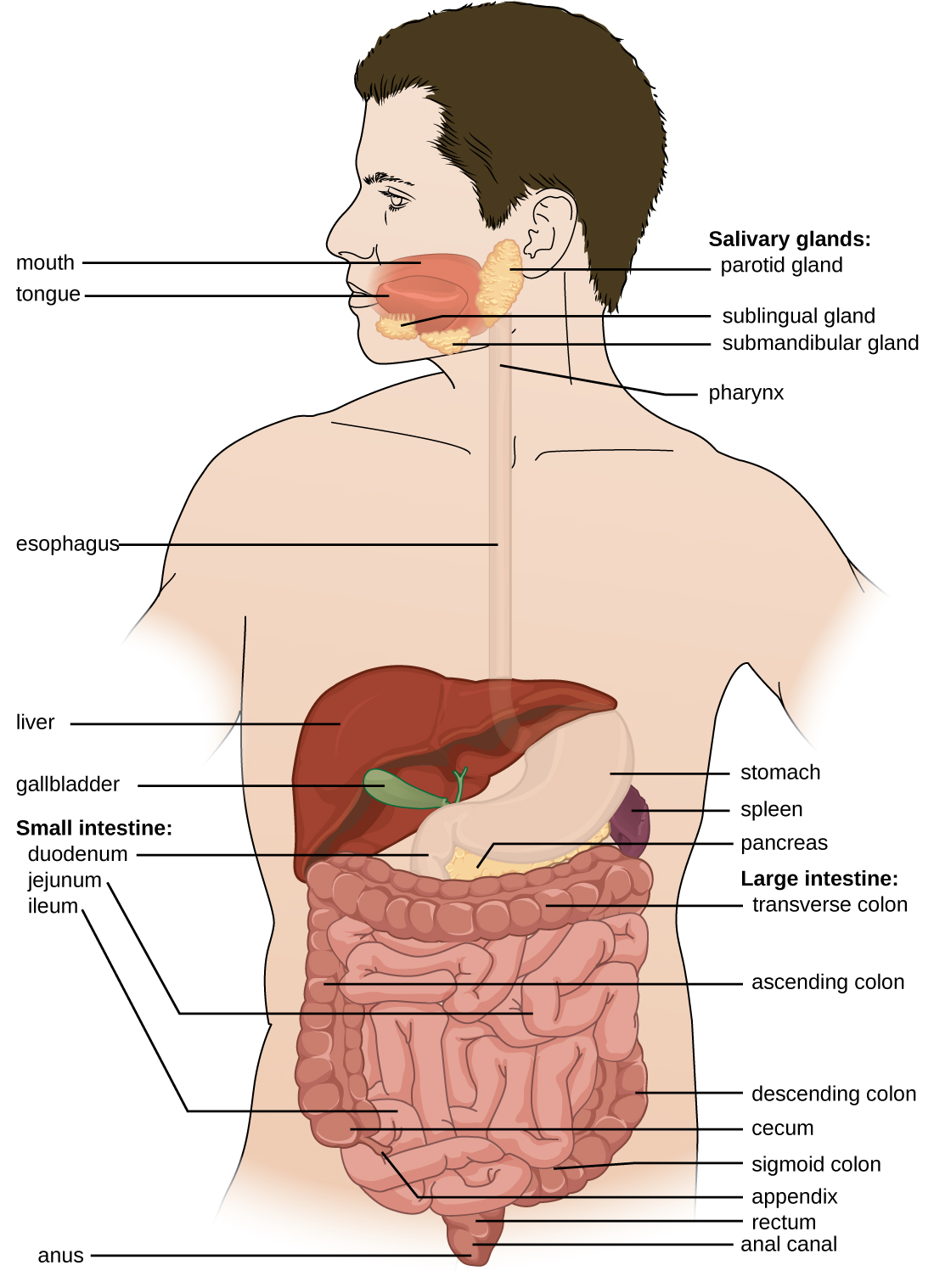
The human digestive system , or the gastrointestinal (GI) tract, begins with the mouth and ends with the anus. The parts of the mouth include the teeth, the gums, the tongue, the oral vestibule (the space between the gums, lips, and teeth), and the oral cavity proper (the space behind the teeth and gums). Other parts of the GI tract are the pharynx, esophagus, stomach, small intestine, large intestine, rectum, and anus ( [link] ). Accessory digestive organs include the salivary glands, liver, gallbladder, spleen, and pancreas.
The digestive system contains normal microbiota, including archaea, bacteria, fungi, protists, and even viruses. Because this microbiota is important for normal functioning of the digestive system, alterations to the microbiota by antibiotics or diet can be harmful. Additionally, the introduction of pathogens to the GI tract can cause infections and diseases. In this section, we will review the microbiota found in a healthy digestive tract and the general signs and symptoms associated with oral and GI infections.
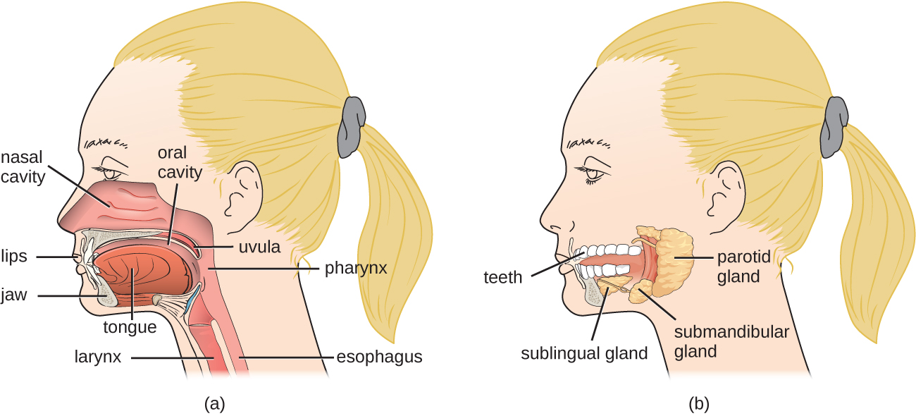
Food enters the digestive tract through the mouth, where mechanical digestion (by chewing) and chemical digestion (by enzymes in saliva ) begin. Within the mouth are the tongue , teeth , and salivary glands , including the parotid, sublingual, and submandibular glands ( [link] ). The salivary glands produce saliva, which lubricates food and contains digestive enzymes.
The structure of a tooth ( [link] ) begins with the visible outer surface, called the crown , which has to be extremely hard to withstand the force of biting and chewing. The crown is covered with enamel , which is the hardest material in the body. Underneath the crown, a layer of relatively hard dentin extends into the root of the tooth around the innermost pulp cavity, which includes the pulp chamber at the top of the tooth and pulp canal, or root canal , located in the root. The pulp that fills the pulp cavity is rich in blood vessels, lymphatic vessels, connective tissue, and nerves. The root of the tooth and some of the crown are covered with cementum , which works with the periodontal ligament to anchor the tooth in place in the jaw bone. The soft tissues surrounding the teeth and bones are called gums , or gingiva . The gingival space or gingival crevice is located between the gums and teeth.
Notification Switch
Would you like to follow the 'Microbiology' conversation and receive update notifications?

|
|

|
|

|
|
|
|
|

|
|

|
|

|
|

|
|

|
|

|