<< Chapter < Page Chapter >> Page >

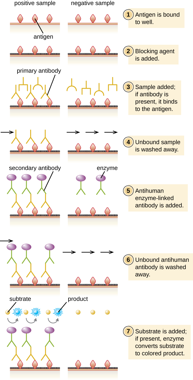
In an indirect ELISA , we quantify antigen-specific antibody rather than antigen. We can use indirect ELISA to detect antibodies against many types of pathogens, including Borrelia burgdorferi ( Lyme disease ) and HIV . There are three important differences between indirect and direct ELISAs as shown in [link] . Rather than using antibody to capture antigen, the indirect ELISA starts with attaching known antigen (e.g., peptides from HIV) to the bottom of the microtiter plate wells. After blocking the unbound sites on the plate, patient serum is added; if antibodies are present ( primary antibody ), they will bind the antigen. After washing away any unbound proteins, the secondary antibody with its conjugated enzyme is directed against the primary antibody (e.g., antihuman immunoglobulin). The secondary antibody allows us to quantify how much antigen-specific antibody is present in the patient’s serum by the intensity of the color produced from the conjugated enzyme-chromogen reaction.

As with several other tests for antibodies discussed in this chapter, there is always concern about cross-reactivity with antibodies directed against some other antigen, which can lead to false-positive results. Thus, we cannot definitively diagnose an HIV infection (or any other type of infection) based on a single indirect ELISA assay. We must confirm any suspected positive test, which is most often done using either an immunoblot that actually identifies the presence of specific peptides from the pathogen or a test to identify the nucleic acids associated with the pathogen, such as reverse transcriptase PCR (RT-PCR) or a nucleic acid antigen test.

a) Diagram of a sandwich ELISA showing what happens in both a positive and a negative sample. First, the primary antibody binds to the well. This is shown as Ys bound to a surface. Next, blocking agent is added. This is shown as a black covering on the surface between the antibodies. Next, the sample is added; if the correct antigen is present it binds to the antibody. In the positive well a circle binds to the antibodies; in the negative well nothing binds to the antibodies. Next, any unbound sample is washed away. Next, antibody-enzyme conjugate is added. This is shown in the positive well as another Y shape that binds to the circles. This new Y shape has a purple circle at the end. These antibodies are also in the negative sample but they are not attached to anything. Next, unbound antibody-enzyme conjugate is washed away; these remain in the positive sample (because they are attached to the antigen) but are washed away in the negative sample. Finally, a substrate is added to both the positive and negative samples. The enzyme in the positive sample turns this substrate blue. b) A plastic plate with many wells. Some are clear, some are blue.
(a) In a sandwich ELISA, a primary antibody is used to first capture an antigen with the primary antibody. A secondary antibody conjugated to an enzyme that also recognizes epitopes on the antigen is added. After the addition of the chromogen, a spectrophotometer measures the absorbance of end product, which is directly proportional to the amount of captured antigen. (b) An ELISA plate shows dilutions of antibodies (left) and antigens (bottom). Higher concentrations result in a darker final color. (credit b: modification of work by U.S. Fish and Wildlife Service Pacific Region)
Diagram of indirect ELISA showing what happens in both a positive and a negative sample. First the antigens are bound to the well. This is shown as diamonds on a surface of both the positive and negative wells. Next, blocking agent is added. This is shown as a black covering on the surface between the antigens. Next, the sample is added. If the correct antibody is present it binds to the antigen. This is shown as Ys in both the positive and negative wells. Otherwise, nothing binds to the antigen. Next, any unbound sample is washed away. In the positive well, there is an antibody bound to the antigen, in the negative well, there is nothing bound to the antigen. Next, anti-human enzyme-linked antibody is added. This is shown as a Ys with a purple circle in both wells. Next, unbound antigen in washed away. In the positive sample this Y remains bound to the old antibody. In the negative well it is no longer present. Finally, substrate is added to both wells. In the positive well, the enzyme changes the substrate to a blue color.
The indirect ELISA is used to quantify antigen-specific antibodies in patient serum for disease diagnosis. Antigen from the suspected disease agent is attached to microtiter plates. The primary antibody comes from the patient’s serum, which is subsequently bound by the enzyme-conjugated secondary antibody. Measuring the production of end product allows us to detect or quantify the amount of antigen-specific antibody present in the patient’s serum.
  • What is the purpose of the secondary antibody in a direct ELISA?
  • What do the direct and indirect ELISAs quantify?

Part 2

Although contacting and testing the 1300 patients for HIV would be time consuming and expensive, administrators hoped to minimize the hospital’s liability by proactively seeking out and treating potential victims of the rogue employee’s crime. Early detection of HIV is important, and prompt treatment can slow the progression of the disease.

There are a variety of screening tests for HIV, but the most widely used is the indirect ELISA. As with other indirect ELISAs, the test works by attaching antigen (in this case, HIV peptides) to a well in a 96-well plate. If the patient is HIV positive, anti-HIV antibodies will bind to the antigen and be identified by the second antibody-enzyme conjugate.

  • How accurate is an indirect ELISA test for HIV, and what factors could impact the test’s accuracy?
  • Should the hospital use any other tests to confirm the results of the indirect ELISA?

Jump to the previous Clinical Focus box. Jump to the next Clinical Focus box.

Get Jobilize Job Search Mobile App in your pocket Now!

Get it on Google Play Download on the App Store Now


Source:  OpenStax, Microbiology. OpenStax CNX. Nov 01, 2016 Download for free at http://cnx.org/content/col12087/1.4
Google Play and the Google Play logo are trademarks of Google Inc.

Notification Switch

Would you like to follow the 'Microbiology' conversation and receive update notifications?

Ask