

| << Chapter < Page | Chapter >> Page > |
In addition to fixation, staining is almost always applied to color certain features of a specimen before examining it under a light microscope. Stains, or dyes, contain salts made up of a positive ion and a negative ion. Depending on the type of dye, the positive or the negative ion may be the chromophore (the colored ion); the other, uncolored ion is called the counterion. If the chromophore is the positively charged ion, the stain is classified as a basic dye ; if the negative ion is the chromophore, the stain is considered an acidic dye .
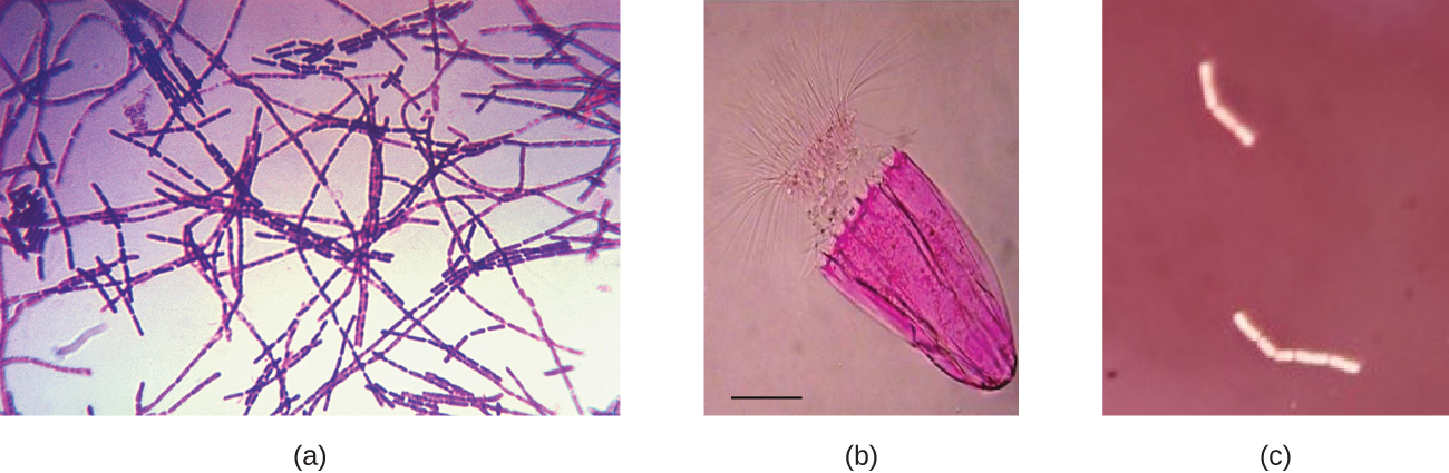
Dyes are selected for staining based on the chemical properties of the dye and the specimen being observed, which determine how the dye will interact with the specimen. In most cases, it is preferable to use a positive stain , a dye that will be absorbed by the cells or organisms being observed, adding color to objects of interest to make them stand out against the background. However, there are scenarios in which it is advantageous to use a negative stain , which is absorbed by the background but not by the cells or organisms in the specimen. Negative staining produces an outline or silhouette of the organisms against a colorful background ( [link] ).
Because cells typically have negatively charged cell walls, the positive chromophores in basic dyes tend to stick to the cell walls, making them positive stains. Thus, commonly used basic dyes such as basic fuchsin , crystal violet , malachite green , methylene blue , and safranin typically serve as positive stains. On the other hand, the negatively charged chromophores in acidic dyes are repelled by negatively charged cell walls, making them negative stains. Commonly used acidic dyes include acid fuchsin , eosin , and rose bengal . [link] provides more detail.
Some staining techniques involve the application of only one dye to the sample; others require more than one dye. In simple staining , a single dye is used to emphasize particular structures in the specimen. A simple stain will generally make all of the organisms in a sample appear to be the same color, even if the sample contains more than one type of organism. In contrast, differential staining distinguishes organisms based on their interactions with multiple stains. In other words, two organisms in a differentially stained sample may appear to be different colors. Differential staining techniques commonly used in clinical settings include Gram staining, acid-fast staining, endospore staining, flagella staining, and capsule staining. [link] provides more detail on these differential staining techniques.
Notification Switch
Would you like to follow the 'Microbiology' conversation and receive update notifications?

|
|

|
|

|
|

|
|

|
|

|
|

|
|
|
|
|

|
|

|