<< Chapter < Page Chapter >> Page >

Like the rest of the plant, the stem has three tissue systems: dermal, vascular, and ground tissue. Each is distinguished by characteristic cell types that perform specific tasks necessary for the plant’s growth and survival.

Dermal tissue

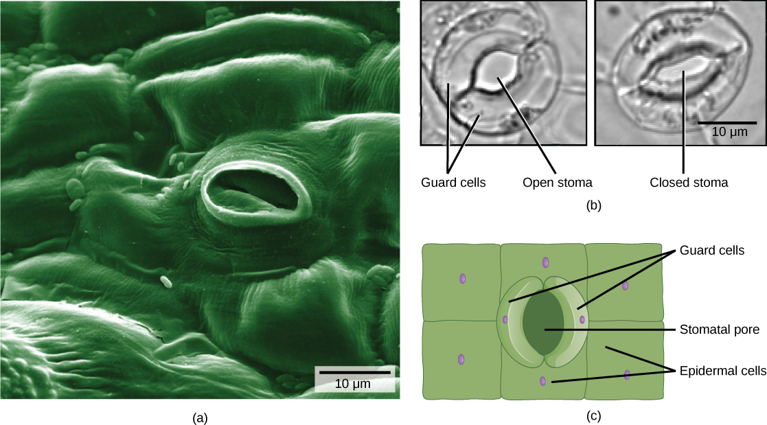
The dermal tissue of the stem consists primarily of epidermis, a single layer of cells covering and protecting the underlying tissue. Woody plants have a tough, waterproof outer layer of cork cells commonly known as bark, which further protects the plant from damage. Epidermal cells are the most numerous and least differentiated of the cells in the epidermis. The epidermis of a leaf also contains openings known as stomata, through which the exchange of gases takes place ( [link] ). Two cells, known as guard cells, surround each leaf stoma, controlling its opening and closing and thus regulating the uptake of carbon dioxide and the release of oxygen and water vapor. Trichomes are hair-like structures on the epidermal surface. They help to reduce transpiration (the loss of water by aboveground plant parts), increase solar reflectance, and store compounds that defend the leaves against predation by herbivores.

 The electron micrograph in part A shows the lumpy, textured of a leaf epidermis. Individual cells look like pillows arranged side by side and fused together. In the center of the image is an oval pore about 10 microns across. Inside the pore, closed guard cells have the appearance of sealed lips. The two light micrographs in part B shows two kidney-shaped guard cells. In the left image, the stoma is open and round. In the right image, the stoma is closed and oval shaped. Part C is an illustration of the leaf epidermis with a oval stomatal pore in the center. Surrounding this pore are two kidney-shaped guard cells. Rectangular epidermal cells surround the guard cells.
Openings called stomata (singular: stoma) allow a plant to take up carbon dioxide and release oxygen and water vapor. The (a) colorized scanning-electron micrograph shows a closed stoma of a eudicot. Each stoma is flanked by two guard cells that regulate its (b) opening and closing. The (c) guard cells sit within the layer of epidermal cells (credit a: modification of work by Louisa Howard, Rippel Electron Microscope Facility, Dartmouth College; credit b: modification of work by June Kwak, University of Maryland; scale-bar data from Matt Russell)

Vascular tissue

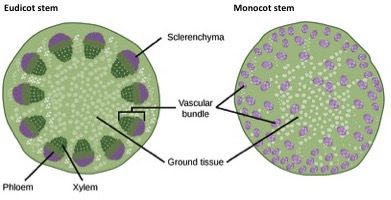
The xylem and phloem that make up the vascular tissue of the stem are arranged in distinct strands called vascular bundles, which run up and down the length of the stem. When the stem is viewed in cross section, the vascular bundles of eudicot stems are arranged in a ring. In plants with stems that live for more than one year, the individual bundles grow together and produce the characteristic growth rings. In monocot stems, the vascular bundles are randomly scattered throughout the ground tissue ( [link] ).

 Part A is cross section of a dicot stem. In the center of the stem is ground tissue. Symmetrically arranged near the outside of the stem are egg-shaped vascular bundles; the narrow end of the egg points inward. The inner part of the vascular bundle is xylem tissue, and the outer part is sclerenchyma tissue. Sandwiched between the xylem and sclerenchyma is the phloem. Part B is a cross section of a monocot stem. In the monocot stem, the vascular bundles are scattered throughout the ground tissue. The bundles are smaller than in the dicot stem, and distinct layers of xylem, phloem and sclerenchyma cannot be discerned.
In (a) eudicot stems, vascular bundles are arranged around the periphery of the ground tissue. The xylem tissue is located toward the interior of the vascular bundle, and phloem is located toward the exterior. Sclerenchyma fibers cap the vascular bundles. In (b) monocot stems, vascular bundles composed of xylem and phloem tissues are scattered throughout the ground tissue.

Xylem tissue has three types of cells: xylem parenchyma, tracheids, and vessel elements. The latter two types conduct water and are dead at maturity. Tracheids are xylem cells with thick secondary cell walls that are lignified. Water moves from one tracheid to another through regions on the side walls known as pits, where secondary walls are absent. Vessel elements are xylem cells with thinner walls; they are shorter than tracheids. Each vessel element is connected to the next by means of a perforation plate at the end walls of the element. Water moves through the perforation plates to travel up the plant.

Get Jobilize Job Search Mobile App in your pocket Now!

Get it on Google Play Download on the App Store Now


Source:  OpenStax, Principles of biology. OpenStax CNX. Aug 09, 2016 Download for free at http://legacy.cnx.org/content/col11569/1.25
Google Play and the Google Play logo are trademarks of Google Inc.

Notification Switch

Would you like to follow the 'Principles of biology' conversation and receive update notifications?

Ask