

| << Chapter < Page | Chapter >> Page > |
The term tissue is used to describe a group of cells found together in the body. These cells work together to perform a certain function. The cells might protect us, fight disease or produce hormones.
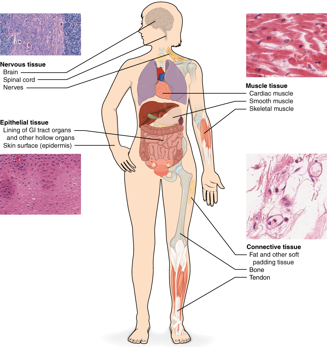
Although there are many types of cells in the human body, they are organized into four broad categories of tissues: epithelial, connective, muscle, and nervous . Each of these categories is characterized by specific functions that contribute to the overall health and maintenance of the body. Histology is the microscopic study of tissue appearance, organization, and function.
Epithelial tissue , also referred to as epithelium, refers to the sheets of cells that cover exterior surfaces of the body, line internal cavities and passageways, and form certain glands. Connective tissue , as its name implies, binds the cells and organs of the body together and functions in the protection, support, and integration of all parts of the body. Muscle tissue is excitable, responding to stimulation and contracting to provide movement. Nervous tissue is also excitable, allowing the formation of signals that communicate between different regions of the body ( [link] ).
The next level of organization is the organ, where several types of tissues come together to form a working unit. Just as knowing the structure and function of cells helps you in your study of tissues, knowledge of tissues will help you understand how organs function. The epithelial and connective tissues are discussed in detail in this chapter. Muscle and nervous tissues will be discussed only briefly in this chapter.
A tissue membrane is a thin layer or sheet of cells that covers the outside of the body (for example, skin), the organs (for example, pericardium), internal passageways that lead to the exterior of the body (for example,inside of cheek), and the lining of the moveable joint cavities. A synovial membrane is a type of connective tissue membrane that lines the cavity of a joint. For example, synovial membranes surround the joints of the shoulder, elbow, and knee which form synovial fluid, a natural lubricant for the joints.
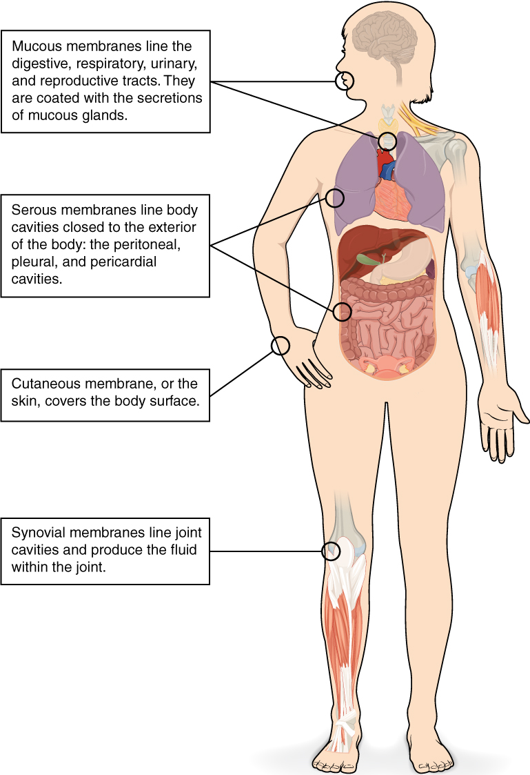
The Cutaneous membrane is composed of epithelium attached to a layer of connective tissue, and is the membrane you know as your skin. The mucous membrane is also made of of connective and epithelial tissues. These membranes line the body cavities and hollow passageways that open to the external environment , and include parts of the digestive, respiratory, excretory, and reproductive tracts. Mucous, produced by glands called goblet cells , covers the membrane.
A serous membrane is an epithelial membrane that lines the closed cavities of the body, that is, those cavities that do not open to the outside. They also cover the organs located within those cavities. Three serous membranes line the thoracic cavity; the two pleura that cover the lungs and the pericardium that covers the heart. A fourth, the peritoneum, is the serous membrane in the abdominal cavity that covers abdominal organs and forms double sheets of mesenteries that suspend many of the digestive organs.
The human body contains more than 200 types of cells that can all be classified into four types of tissues: epithelial, connective, muscle, and nervous. Epithelial tissues act as coverings controlling the movement of materials across the surface. Connective tissue integrates the various parts of the body and provides support and protection to organs. Muscle tissue allows the body to move. Nervous tissues send information from one part of the body to another.
Different types of tissues form membranes that enclose organs, provide a friction-free interaction between organs, and keep organs together. Synovial membranes are connective tissue membranes that protect and line the joints. Epithelial membranes are formed from epithelial tissue attached to a layer of connective tissue. There are three types of epithelial membranes: mucous, which contain glands; serous, which secrete fluid; and cutaneous which makes up the skin.
Notification Switch
Would you like to follow the 'Histology' conversation and receive update notifications?

|
|

|
|

|
|

|
|

|
|

|
|

|
|

|
|

|
|

|