<< Chapter < Page Chapter >> Page >

Sponges reproduce both sexually and asexually. Asexual reproduction is either by fragmentation    (in which a piece of the sponge breaks off and develops into a new individual), or budding    (an outgrowth from the parent that eventually detaches). A type of asexual reproduction found only in freshwater sponges occurs through the formation of gemmules , clusters of cells surrounded by a tough outer layer. Gemmules survive hostile environments and can attach to a substrate and grow into a new sponge.

Sponges are monoecious    (or hermaphroditic), meaning one individual can produce both eggs and sperm. Sponges may be sequentially hermaphroditic, producing eggs first and sperm later. Eggs arise from amoebocytes and are retained within the spongocoel, whereas sperm arise from choanocytes and are ejected through the osculum. Sperm carried by water currents fertilize the eggs of other sponges. Early larval development occurs within the sponge, and free-swimming larvae are then released through the osculum. This is the only time that sponges exhibit mobility. Sponges are sessile as adults and spend their lives attached to a fixed substrate.

Concept in action

Watch this video that demonstrates the feeding of sponges.

Cnidarians

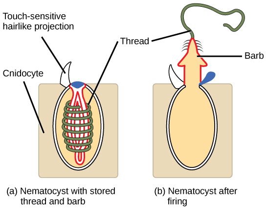
The phylum Cnidaria    includes animals that show radial or biradial symmetry and are diploblastic. Nearly all (about 99 percent) cnidarians are marine species. Cnidarians have specialized cells known as cnidocytes (“stinging cells”) containing organelles called nematocysts . These cells are concentrated around the mouth and tentacles of the animal and can immobilize prey with toxins. Nematocysts contain coiled threads that may bear barbs. The outer wall of the cell has a hairlike projection that is sensitive to touch. When touched, the cells fire the toxin-containing coiled threads that can penetrate and stun the predator or prey (see [link] ).

The illustration shows a nematocyst before (a) and after (b) firing. The nematocyst is a large, oval organelle inside a rectangular cnidocyte cell. The nematocyst is flush with the plasma membrane, and a touch-sensitive hairlike projection extends from the nematocyst to the cell’s exterior. Inside the nematocyst, a thread is coiled around an inverted barb. Upon firing, a lid on the nematocyst opens. The barb pops out of the cell and the thread uncoils.
Animals from the phylum Cnidaria have stinging cells called cnidocytes. Cnidocytes contain large organelles called (a) nematocysts that store a coiled thread and barb. When hairlike projections on the cell surface are touched, (b) the thread, barb, and a toxin are fired from the organelle.

Cnidarians display two distinct body plans: polyp    or “stalk” and medusa    or “bell” ( [link] ). Examples of the polyp form are freshwater species of the genus Hydra ; perhaps the best-known medusoid animals are the jellies (jellyfish). Polyps are sessile as adults, with a single opening to the digestive system (the mouth) facing up with tentacles surrounding it. Medusae are motile, with the mouth and tentacles hanging from the bell-shaped body. In other cnidarians, both a polyp and medusa form exist, and the life cycle alternates between these forms.

The illustration compares the medusa (a) and polyp (b) body plans. The medusa is dome-shaped, with tentacle-like appendages hanging down from the edges of the dome. The polyp looks like a tree, with a trunk at the bottom and branches at the top. Both the medusa and polyp have two tissue layers, with mesoglea in between. The mesoglea is thicker in the dome of the medusa than in the polyp. Both also have a central body cavity.
Cnidarians have two distinct body plans, the (a) medusa and the (b) polyp. All cnidarians have two tissue layers, with a jelly-like mesoglea between them.

Physiological processes of cnidarians

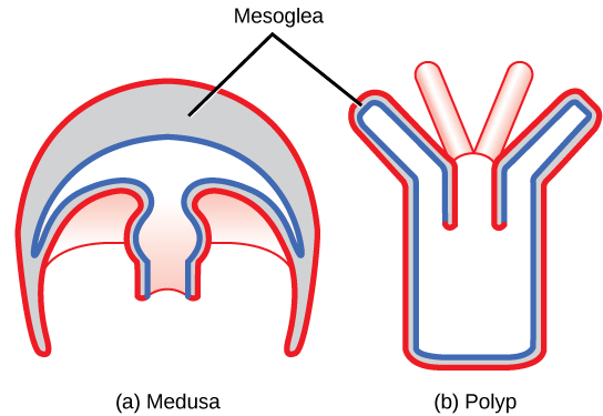
All cnidarians have two tissue layers. The outer layer is called the epidermis    , whereas the inner layer is called the gastrodermis    and lines the digestive cavity. Between these two layers is a non-living, jelly-like mesoglea    . There are differentiated cell types in each tissue layer, such as nerve cells, enzyme-secreting cells, and nutrient-absorbing cells, as well as intercellular connections between the cells. However, organs and organ systems are not present in this phylum.

Questions & Answers

what is biology
Hajah Reply
the study of living organisms and their interactions with one another and their environments
AI-Robot
what is biology
Victoria Reply
HOW CAN MAN ORGAN FUNCTION
Alfred Reply
the diagram of the digestive system
Assiatu Reply
allimentary cannel
Ogenrwot
How does twins formed
William Reply
They formed in two ways first when one sperm and one egg are splited by mitosis or two sperm and two eggs join together
Oluwatobi
what is genetics
Josephine Reply
Genetics is the study of heredity
Misack
how does twins formed?
Misack
What is manual
Hassan Reply
discuss biological phenomenon and provide pieces of evidence to show that it was responsible for the formation of eukaryotic organelles
Joseph Reply
what is biology
Yousuf Reply
the study of living organisms and their interactions with one another and their environment.
Wine
discuss the biological phenomenon and provide pieces of evidence to show that it was responsible for the formation of eukaryotic organelles in an essay form
Joseph Reply
what is the blood cells
Shaker Reply
list any five characteristics of the blood cells
Shaker
lack electricity and its more savely than electronic microscope because its naturally by using of light
Abdullahi Reply
advantage of electronic microscope is easily and clearly while disadvantage is dangerous because its electronic. advantage of light microscope is savely and naturally by sun while disadvantage is not easily,means its not sharp and not clear
Abdullahi
cell theory state that every organisms composed of one or more cell,cell is the basic unit of life
Abdullahi
is like gone fail us
DENG
cells is the basic structure and functions of all living things
Ramadan
What is classification
ISCONT Reply
is organisms that are similar into groups called tara
Yamosa
in what situation (s) would be the use of a scanning electron microscope be ideal and why?
Kenna Reply
A scanning electron microscope (SEM) is ideal for situations requiring high-resolution imaging of surfaces. It is commonly used in materials science, biology, and geology to examine the topography and composition of samples at a nanoscale level. SEM is particularly useful for studying fine details,
Hilary
cell is the building block of life.
Condoleezza Reply
Got questions? Join the online conversation and get instant answers!
Jobilize.com Reply

Get Jobilize Job Search Mobile App in your pocket Now!

Get it on Google Play Download on the App Store Now


Source:  OpenStax, Concepts of biology. OpenStax CNX. Feb 29, 2016 Download for free at http://cnx.org/content/col11487/1.9
Google Play and the Google Play logo are trademarks of Google Inc.

Notification Switch

Would you like to follow the 'Concepts of biology' conversation and receive update notifications?

Ask