

| << Chapter < Page | Chapter >> Page > |
he wall of the stomach is made of the same four layers as most of the rest of the alimentary canal, but with adaptations to the mucosa and muscularis for the unique functions of this organ. In addition to the typical circular and longitudinal smooth muscle layers, the muscularis has an third layer of muscle, the oblique muscle layer ( [link] )which allows the stomach to vigorously churn food, mechanically breaking it down into smaller particles.
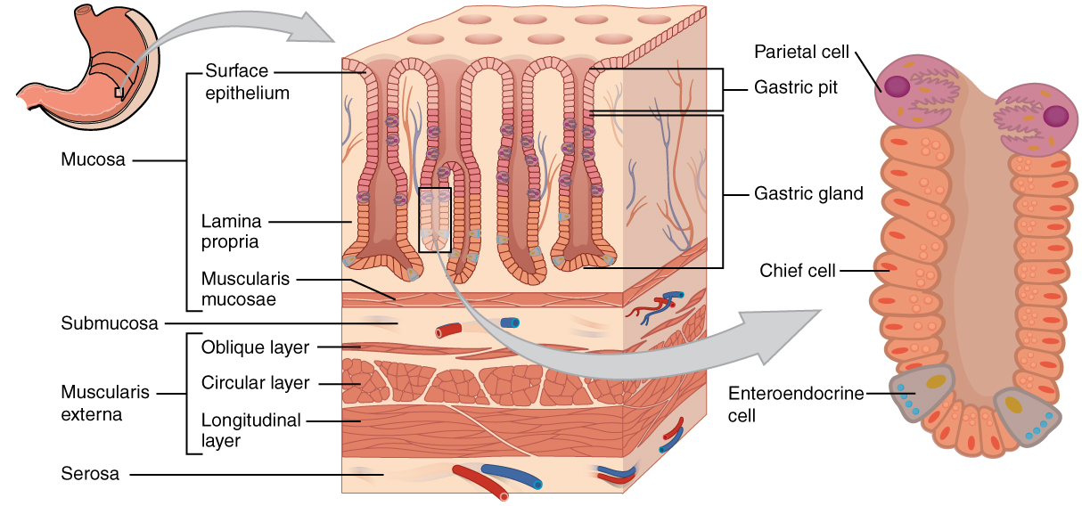
The stomach mucosa’s epithelial lining consists only of surface mucus cells, which secrete a protective coat of alkaline mucus. A vast number of gastric pits dot the surface of the epithelium, giving it the appearance of a well-used pincushion, and mark the entry to each gastric gland , which secretes a complex digestive fluid referred to as gastric juice .
Although the walls of the gastric pits are made up primarily of mucus cells, the gastric glands also include chief cells and parietal cells. Cells that make up the area of the pyloris secrete mucus and a number of hormones, including the majority of the stimulatory hormone, gastrin . The much larger glands of the fundus and body of the stomach, the site of most chemical digestion, produce most of the gastric secretions. These glands are made up of a variety of secretory cells. These include parietal cells, chief cells, mucous neck cells, and enteroendocrine cells.
Parietal cells —Located primarily in the middle region of the gastric glands are parietal cells which produce both hydrochloric acid (HCl) and intrinsic factor . HCl is responsible for the high acidity (pH 1.5 to 3.5) of the stomach contents and is needed to activate the protein-digesting enzyme, pepsin . The acidity also kills much of the bacteria you ingest with food and helps to break down proteins, making them more available for enzymatic digestion. Intrinsic factor is a glycoprotein necessary for the absorption of vitamin B 12 in the small intestine.
Chief cells —Also located primarily in the gastric glands are chief cells , which secrete pepsinogen , the inactive substance that will convert to pepsin. HCl is necessary for the conversion of pepsinogen to pepsin. Enteroendocrine cells —Finally, enteroendocrine cells which are also found in the gastric glands secrete various hormones. Do not take notes on table below.
[link] describes the digestive functions of important hormones secreted by the stomach.
| Hormones Secreted by the Stomach | ||||
|---|---|---|---|---|
| Hormone | Production site | Production stimulus | Target organ | Action |
| Gastrin | Stomach mucosa, mainly G cells of the pyloric antrum | Presence of peptides and amino acids in stomach | Stomach | Increases secretion by gastric glands; promotes gastric emptying |
| Gastrin | Stomach mucosa, mainly G cells of the pyloric antrum | Presence of peptides and amino acids in stomach | Small intestine | Promotes intestinal muscle contraction |
| Gastrin | Stomach mucosa, mainly G cells of the pyloric antrum | Presence of peptides and amino acids in stomach | Ileocecal valve | Relaxes valve |
| Gastrin | Stomach mucosa, mainly G cells of the pyloric antrum | Presence of peptides and amino acids in stomach | Large intestine | Triggers mass movements |
| Ghrelin | Stomach mucosa, mainly fundus | Fasting state (levels increase just prior to meals) | Hypothalamus | Regulates food intake, primarily by stimulating hunger and satiety |
| Histamine | Stomach mucosa | Presence of food in the stomach | Stomach | Stimulates parietal cells to release HCl |
| Serotonin | Stomach mucosa | Presence of food in the stomach | Stomach | Contracts stomach muscle |
| Somatostatin | Mucosa of stomach, especially pyloric antrum; also duodenum | Presence of food in the stomach; sympathetic axon stimulation | Stomach | Restricts all gastric secretions, gastric motility, and emptying |
| Somatostatin | Mucosa of stomach, especially pyloric antrum; also duodenum | Presence of food in the stomach; sympathetic axon stimulation | Pancreas | Restricts pancreatic secretions |
| Somatostatin | Mucosa of stomach, especially pyloric antrum; also duodenum | Presence of food in the stomach; sympathetic axon stimulation | Small intestine | Reduces intestinal absorption by reducing blood flow |
Notification Switch
Would you like to follow the 'Digestive system' conversation and receive update notifications?

|
|

|
|

|
|

|
|

|
|

|
|

|
|

|
|
|
|
|

|