<< Chapter < Page Chapter >> Page >
C1s high resolution XP spectra of graphite, nanodiamond, and graphite samples with increasing laser power treatment. Adapted from P. Merel, M. Tabbal, M. Chaker, S. Moisa, and J. Margot, Appl. Surf. Sci. , 1998, 136 , 105.

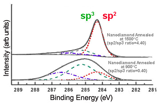
Alternatively, annealing nanodiamond thin films at very high temperatures creates graphitic layers on the nanodiamond surface, increasing sp 2 content. The extent of graphitization increases with the temperature at which the sample is annealed, as shown in [link] .

Deconvoluted high resolution C1s XP spectra for annealed nanodiamond. Adapted from F. Y. Xie, W. G. Xie, J. Chen, X. Liu, D. Y. Lu, and W. H. Zhang, J. Vac. Sci. Tech. B , 2008, 26 , 102.

Reaction completion

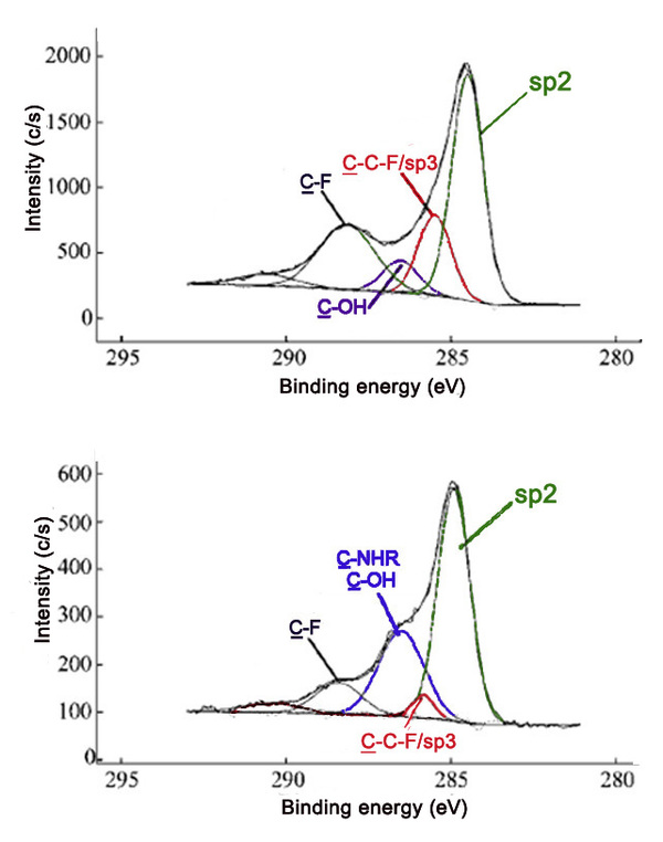
Comparing the relative intensities of various C1s peaks can be powerful in verifying that a reaction has occurred. Fluorinated carbon materials are often used as precursors to a broad range of variously functionalized materials. Reaction of fluorinated SWNTs (F-SWNTs) with polyethyleneimine (PEI) leads to decreases in the covalent carbon-fluoride C1s peak, as well as the evolution of the amine C1s peak. These changes are observed in the C1s spectra of the two samples ( [link] ).

High resolution C1s XP spectra of F-SWNTs (top) and PEI-SWNTs (bottom). Adapted with permission from E. P. Dillon, C. A. Crouse, and A. R. Barron, ACS Nano , 2008, 2 , 156. Copyright: American Chemical Society (2008).

Nature and extent of functionalization

XPS can also be applied to determine the nature and extent of functionalization. In general, binding energy increases with decreasing electron density about the atom. Species with more positive oxidation states have higher binding energies, while more reduced species experience a greater degree of shielding, thus increasing the ease of electron removal.

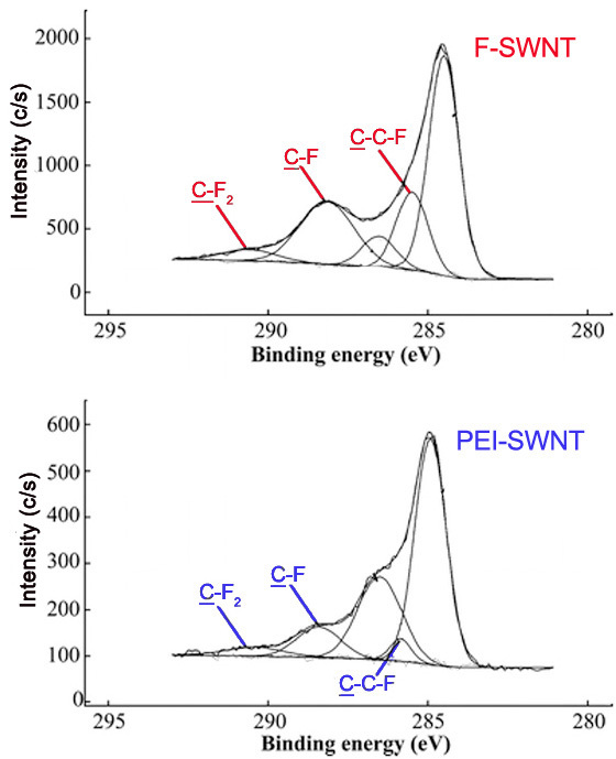
The method of fluorination of carbon materials and such factors as temperature and length of fluorination affect the extent of fluoride addition as well as the types of carbon-fluorine bonds present. A survey scan can be used to determine the amount of fluorine compared to carbon. High resolution scans of the C1s and F1s peaks can also give information about the proportion and types of bonds. A shift in the peaks, as well as changes in peak width and intensity, can be observed in spectra as an indication of fluorination of graphite. [link] shows the Cls and F1s spectra of samples containing varying ratios of carbon to fluorine.

C1s and F1s high resolution XP spectra for graphite fluorides. Adapted from I. Palchan, M. Crespin, H. Estrade-Szwarckopf, and B. Rousseau. Chem. Phys. Lett. , 1989, 157 , 321.

Furthermore, different carbon-fluorine bonds show characteristic peaks in high resolution C1s and F1s spectra. The carbon-fluorine interactions in a material can range from ionic to covalent. Covalent carbon-fluorine bonds show higher core electron binding energies than bonds more ionic in character. The method of fluorination affects the nature of the fluorine bonds. Graphite intercalation compounds are characterized by ionic carbon-fluorine bonding. [link] shows the F1s spectra for two fluorinated exfoliated graphite samples prepared with different methods.

High resolution F1s XP spectra of two fluorinated exfoliated graphite samples. Adapted from A. Tressaud, F. Moguet, S. Flandrois, M. Chambon, C. Guimon, G. Nanse, E. Papirer, V. Gupta, and O.P. Bahl. J. Phys. Chem. Solids , 1996, 57 , 745.

Also, the peaks for carbons attached to a single fluorine atom, two fluorine atoms, and carbons attached to fluorines have characteristic binding energies. These peaks are seen in that C1s spectra of F- and PEI-SWNTs shown in [link] .

High resolution C1s XP spectra of F-SWNTs (top) and PEI-SWNTs (bottom). Adapted with permission from E. P. Dillon, C. A. Crouse, and A. R. Barron, ACS Nano , 2008, 2 , 156. Copyright: American Chemical Society (2008).

[link] lists various bonds and functionalities and the corresponding C1s binding energies, which may be useful in assigning peaks in a C1s spectrum, and consequently in characterizing the surface of a material.

Summary of selected C1s binding energies.
Bond/Group Binding Energy (eV)
C-C 284.0-286.0
C-C (sp 2 ) 284.3-284.6
C-C (sp 3 ) 285.0-286.0
C-N 285.2-288.4
C-NR 2 (amine) 285.5-286.4
O=C-NH (amide) 287.9-288.6
-C = N (nitrile) 266.3-266.8
C-O 286.1-290.0
O=C-OH (carboxyl) 288.0-289.4
-C-O (epoxy) 286.1-287.1
-C-OH (hydroxyl) 286.4-286.7
-C-O-C- (ether) 286.1-288.0
-C=O (aldehyde/ketone) 287.1-288.1
C-F 287.0-293.4
-C-F (covalent) 287.7-290.2
-C-F (ionic) 287.0-287.4
C -C-F 286.0-287.7
C-F 2 291.6-292.4
C-F 3 292.4-293.4
C-S 285.2-287.5
C-Cl 287.0-287.2

Conclusion

X-ray photoelectron spectroscopy is a facile and effective method for determining the elemental composition of a material’s surface. As a quantitative method, it gives the relative ratios of detectable elements on the surface of the material. Additional analysis can be done to further elucidate the surface structure. Hybridization, bonding, functionalities, and reaction progress are among the characteristics that can be inferred using XPS. The application of XPS to carbon nanomaterials provides much information about the material, particularly the first few atomic layers, which are most important for the properties and uses of carbon nanomaterials.

Bibliography

  • J. Bockris, Modern Electrochemistry 2A , 2 nd ed., Springer (2001).
  • E. P. Dillon, C. A. Crouse, and A. R. Barron, ACS Nano , 2008, 2 , 156.
  • Y. G. Gogotsi, and I. V. Uvarova, Nanostructured materials and coatings for biomedical and sensor applications, Kluwer Academic Publishing (2003).
  • F. Liang, J. M. Beach, P. K. Rai, W. H. Guo, R. H. Hauge, M. Pasquali, R. E. Smalley, and W. E. Billups, Chem. Mater. , 2006, 18 , 1520.
  • P. Merel, M. Tabbal, M. Chaker, S. Moisa, and J. Margot, Appl. Surf. Sci. , 1998, 136 , 105.
  • T. Nakajima, Graphite fluorides and carbon-fluorine compounds , CRC Press, Boca Raton (1991).
  • I. Palhan, M. Crespin, H. Estrade-Szwarckopf, and B. Rousseau. Chem. Phys. Lett. , 1989, 157 , 321.
  • A. Tressaud, F. Moguet, S. Flandrois, M. Chambon, C. Guimon, G. Nanse, E. Papirer, V. Gupta, and O. P. Bahl. J. Phys. Chem. Solids , 1996, 57 , 745.
  • F. Y. Xie, W. G. Xie, J. Chen, X. Liu, D. Y. Lu, and W. H. Zhang, J. Vac. Sci. Tech. B , 2008, 26 , 102.
  • C. M. Yang, H. Kanoh, K. Kaneko, M. Yudasaka, and S. Iijima, J. Phys. Chem. B , 2002, 106 , 8994.

Questions & Answers

what is biology
Hajah Reply
the study of living organisms and their interactions with one another and their environments
AI-Robot
what is biology
Victoria Reply
HOW CAN MAN ORGAN FUNCTION
Alfred Reply
the diagram of the digestive system
Assiatu Reply
allimentary cannel
Ogenrwot
How does twins formed
William Reply
They formed in two ways first when one sperm and one egg are splited by mitosis or two sperm and two eggs join together
Oluwatobi
what is genetics
Josephine Reply
Genetics is the study of heredity
Misack
how does twins formed?
Misack
What is manual
Hassan Reply
discuss biological phenomenon and provide pieces of evidence to show that it was responsible for the formation of eukaryotic organelles
Joseph Reply
what is biology
Yousuf Reply
the study of living organisms and their interactions with one another and their environment.
Wine
discuss the biological phenomenon and provide pieces of evidence to show that it was responsible for the formation of eukaryotic organelles in an essay form
Joseph Reply
what is the blood cells
Shaker Reply
list any five characteristics of the blood cells
Shaker
lack electricity and its more savely than electronic microscope because its naturally by using of light
Abdullahi Reply
advantage of electronic microscope is easily and clearly while disadvantage is dangerous because its electronic. advantage of light microscope is savely and naturally by sun while disadvantage is not easily,means its not sharp and not clear
Abdullahi
cell theory state that every organisms composed of one or more cell,cell is the basic unit of life
Abdullahi
is like gone fail us
DENG
cells is the basic structure and functions of all living things
Ramadan
What is classification
ISCONT Reply
is organisms that are similar into groups called tara
Yamosa
in what situation (s) would be the use of a scanning electron microscope be ideal and why?
Kenna Reply
A scanning electron microscope (SEM) is ideal for situations requiring high-resolution imaging of surfaces. It is commonly used in materials science, biology, and geology to examine the topography and composition of samples at a nanoscale level. SEM is particularly useful for studying fine details,
Hilary
cell is the building block of life.
Condoleezza Reply
Got questions? Join the online conversation and get instant answers!
Jobilize.com Reply

Get Jobilize Job Search Mobile App in your pocket Now!

Get it on Google Play Download on the App Store Now


Source:  OpenStax, Nanomaterials and nanotechnology. OpenStax CNX. May 07, 2014 Download for free at http://legacy.cnx.org/content/col10700/1.13
Google Play and the Google Play logo are trademarks of Google Inc.

Notification Switch

Would you like to follow the 'Nanomaterials and nanotechnology' conversation and receive update notifications?

Ask