<< Chapter < Page Chapter >> Page >

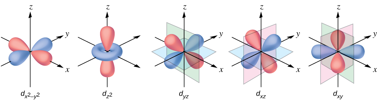
The other common geometry is square planar. It is possible to consider a square planar geometry as an octahedral structure with a pair of trans ligands removed. The removed ligands are assumed to be on the z -axis. This changes the distribution of the d orbitals, as orbitals on or near the z -axis become more stable, and those on or near the x- or y -axes become less stable. This results in the octahedral t 2 g and the e g sets splitting and gives a more complicated pattern with no simple Δ oct . The basic pattern is:

A diagram is shown with four rows of vertically oriented rectangles. The lower level has two rectangles with a space between them. The rectangle on the left is labeled, “d subscript x z,” below. The rectangle to its right is similarly labeled, “d subscript y z.” Just above, the second row contains only 1 rectangle above and between the lower two. This rectangle is labeled, “d subscript z squared.” Just above, the third row contains only 1 rectangle directly above. This rectangle is labeled, “d subscript x z.” Just above, the fourth row contains only 1 rectangle directly above. This rectangle is labeled, “d subscript x squared minus y squared.”

Magnetic moments of molecules and ions

Experimental evidence of magnetic measurements supports the theory of high- and low-spin complexes. Remember that molecules such as O 2 that contain unpaired electrons are paramagnetic. Paramagnetic substances are attracted to magnetic fields. Many transition metal complexes have unpaired electrons and hence are paramagnetic. Molecules such as N 2 and ions such as Na + and [Fe(CN) 6 ] 4− that contain no unpaired electrons are diamagnetic. Diamagnetic substances have a slight tendency to be repelled by magnetic fields.

When an electron in an atom or ion is unpaired, the magnetic moment due to its spin makes the entire atom or ion paramagnetic. The size of the magnetic moment of a system containing unpaired electrons is related directly to the number of such electrons: the greater the number of unpaired electrons, the larger the magnetic moment. Therefore, the observed magnetic moment is used to determine the number of unpaired electrons present. The measured magnetic moment of low-spin d 6 [Fe(CN) 6 ] 4− confirms that iron is diamagnetic, whereas high-spin d 6 [Fe(H 2 O) 6 ] 2+ has four unpaired electrons with a magnetic moment that confirms this arrangement.

Colors of transition metal complexes

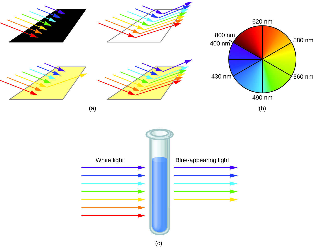
When atoms or molecules absorb light at the proper frequency, their electrons are excited to higher-energy orbitals. For many main group atoms and molecules, the absorbed photons are in the ultraviolet range of the electromagnetic spectrum, which cannot be detected by the human eye. For coordination compounds, the energy difference between the d orbitals often allows photons in the visible range to be absorbed.

The human eye perceives a mixture of all the colors, in the proportions present in sunlight, as white light. Complementary colors, those located across from each other on a color wheel, are also used in color vision. The eye perceives a mixture of two complementary colors, in the proper proportions, as white light. Likewise, when a color is missing from white light, the eye sees its complement. For example, when red photons are absorbed from white light, the eyes see the color green. When violet photons are removed from white light, the eyes see lemon yellow. The blue color of the [Cu(NH 3 ) 4 ] 2+ ion results because this ion absorbs orange and red light, leaving the complementary colors of blue and green ( [link] ).

This figure includes three diagrams. In a, red, orange, yellow, green, blue, indigo, and violet incident arrows point from the upper left to the lower right toward one black, one white, and two yellow rectangular surfaces. No arrows are indicated leaving the black surface. On the white surface, arrows of all included colors extend from the surface of the rectangle extending from just right of the tips of the incident arrows that approach the rectangle to the upper right. For the first yellow surface, only a yellow arrow extends from a point just past the tip of the yellow incident arrow to the upper right. For the second yellow surface, all colors of arrows except indigo extend from points just past the tips of the incident arrows to the upper right. In b, a circle shaded in red at the upper left blends to orange, yellow, green, blue, indigo, and violet moving clockwise about the circle. The leftmost side of the red region has a radius that is labeled, “800 n m,” at the edge of the circle. A radius drawn to the top of the circle in the red-orange region is labeled, “620 n m.” A radius drawn to a point near the center of the first quadrant of the circle in the orange-yellow region is labeled, “580 n m.” A radius drawn to a point near the center of the second quadrant of the circle in the yellow-green region is labeled, “560 n m.” A radius drawn to the bottom of the circle in the blue region is labeled, “490 n m.” A radius drawn to a point near the center of the third quadrant of the circle in the indigo region is labeled, “430 n m.” An unlabeled radius is drawn to the leftmost point on the circle in the violet region. The violet region ends where the red region began on the circle. This radius is labeled, “400 n m,” just to the left and below the, “800 n m,” label associated with the red region. In c, a test tube containing a blue substance is shown. To the left of the test tube, incident colored arrows are shown pointing to the test tube. The colors of the arrows in order from bottom to top are red, orange, yellow, green, blue, indigo, and violet. Above these arrows is the label, “White light.” To the right of the test tube, yellow, green, blue, indigo, and violet arrows point right. These arrows are positioned at the same level on the test tube as their matching incident arrows on the left of the test tube. Above these arrows is the label, “Blue-appearing light.”
(a) An object is black if it absorbs all colors of light. If it reflects all colors of light, it is white. An object has a color if it absorbs all colors except one, such as this yellow strip. The strip also appears yellow if it absorbs the complementary color from white light (in this case, indigo). (b) Complementary colors are located directly across from one another on the color wheel. (c) A solution of [Cu(NH 3 ) 4 ] 2+ ions absorbs red and orange light, so the transmitted light appears as the complementary color, blue.

Questions & Answers

what is biology
Hajah Reply
the study of living organisms and their interactions with one another and their environments
AI-Robot
what is biology
Victoria Reply
HOW CAN MAN ORGAN FUNCTION
Alfred Reply
the diagram of the digestive system
Assiatu Reply
allimentary cannel
Ogenrwot
How does twins formed
William Reply
They formed in two ways first when one sperm and one egg are splited by mitosis or two sperm and two eggs join together
Oluwatobi
what is genetics
Josephine Reply
Genetics is the study of heredity
Misack
how does twins formed?
Misack
What is manual
Hassan Reply
discuss biological phenomenon and provide pieces of evidence to show that it was responsible for the formation of eukaryotic organelles
Joseph Reply
what is biology
Yousuf Reply
the study of living organisms and their interactions with one another and their environment.
Wine
discuss the biological phenomenon and provide pieces of evidence to show that it was responsible for the formation of eukaryotic organelles in an essay form
Joseph Reply
what is the blood cells
Shaker Reply
list any five characteristics of the blood cells
Shaker
lack electricity and its more savely than electronic microscope because its naturally by using of light
Abdullahi Reply
advantage of electronic microscope is easily and clearly while disadvantage is dangerous because its electronic. advantage of light microscope is savely and naturally by sun while disadvantage is not easily,means its not sharp and not clear
Abdullahi
cell theory state that every organisms composed of one or more cell,cell is the basic unit of life
Abdullahi
is like gone fail us
DENG
cells is the basic structure and functions of all living things
Ramadan
What is classification
ISCONT Reply
is organisms that are similar into groups called tara
Yamosa
in what situation (s) would be the use of a scanning electron microscope be ideal and why?
Kenna Reply
A scanning electron microscope (SEM) is ideal for situations requiring high-resolution imaging of surfaces. It is commonly used in materials science, biology, and geology to examine the topography and composition of samples at a nanoscale level. SEM is particularly useful for studying fine details,
Hilary
cell is the building block of life.
Condoleezza Reply
Got questions? Join the online conversation and get instant answers!
Jobilize.com Reply

Get Jobilize Job Search Mobile App in your pocket Now!

Get it on Google Play Download on the App Store Now


Source:  OpenStax, Chemistry. OpenStax CNX. May 20, 2015 Download for free at http://legacy.cnx.org/content/col11760/1.9
Google Play and the Google Play logo are trademarks of Google Inc.

Notification Switch

Would you like to follow the 'Chemistry' conversation and receive update notifications?

Ask