<< Chapter < Page Chapter >> Page >

Endochondral ossification

Image A shows a small piece of hyaline cartilage that looks like a bone but without the characteristic enlarged ends. The hyaline cartilage is surrounded by a thin perichondrium. In image B, the hyaline cartilage has increased in size and the ends have begun to bulge outwards. A group of dark granules form at the center of the cartilage. This is labeled the calcified matrix, as opposed to the rest of the cartilage, which is uncalcified matrix. In image C, the hyaline cartilage has again increased in size and spongy bone has formed at the calcified matrix. This is now called the primary ossification center. A nutrient artery has invaded the ossification center and is growing through the cavities of the new spongy bone. In image D, the cartilage now looks like a bone, as it has greatly increased in size and each end has two bulges. Only the proximal half of the bone is shown in all of the remaining images. In image D, spongy bone has completely developed in the medullary cavity, which is surrounded, on both sides, by compact bone. Now, the calcified matrix is located at the border between the proximal metaphysis and the proximal epiphysis. The epiphysis is still composed of uncalcified matrix. In image E, arteries and veins have now invaded the epiphysis, forming a calcified matrix at its center. This is called a secondary ossification center. In image F, the interior of the epiphysis is now completely calcified into bone. The outer edge of the epiphysis remains as cartilage, forming the articular cartilage at the joint. In addition, the border between the epiphysis and the metaphysis remains uncalcified, forming the epiphyseal plate.
Endochondral ossification follows five steps. (a) Mesenchymal cells differentiate into chondrocytes. (b) The cartilage model of the future bony skeleton and the perichondrium form. (c) Capillaries penetrate cartilage. Perichondrium transforms into periosteum. Periosteal collar develops. Primary ossification center develops. (d) Cartilage and chondrocytes continue to grow at ends of the bone. (e) Secondary ossification centers develop. (f) Cartilage remains at epiphyseal (growth) plate and at joint surface as articular cartilage.

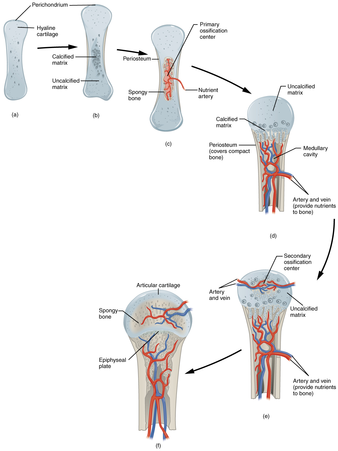
The chondrocytes in the center of the cartilaginous model grow in size. As the matrix calcifies, nutrients can no longer reach the chondrocytes. This results in their death and the disintegration of the surrounding cartilage. Blood vessels invade the resulting spaces, not only enlarging the cavities but also carrying osteogenic cells with them, many of which will become osteoblasts. These enlarging spaces eventually combine to become the medullary cavity.

How bones grow in length

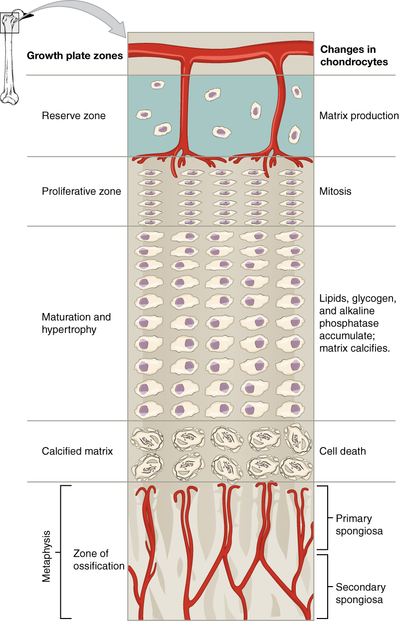
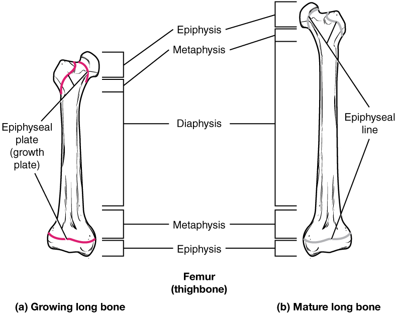
The epiphyseal plate is the area of growth in a long bone. It is a layer of hyaline cartilage where ossification occurs in immature bones. On the epiphyseal side of the epiphyseal plate, cartilage is formed. On the diaphyseal side, cartilage is ossified, and the diaphysis grows in length.

Longitudinal bone growth

This illustration shows the zones bordering the epiphyseal plate of the epiphysis. The topmost layer of the epiphysis is the reserve zone, which is colored blue because it is made of cartilage. Two arteries are shown travelling through this zone to supply nutrients to the second zone: the proliferative zone. Here, five chondrocytes are undergoing mitosis. They continually divide, producing five long rows of chondrocytes. The next zone is the zone of maturation and hypertrophy. Here, lipids, glycogen and alkaline phosphatase accumulate, causing the cartilaginous matrix to calcify. This zone consists of five rows of ten chondrocytes which are increasing in size as one moves down a row. The next zone is the calcified matrix. Here, the chondrocytes have hardened and die as the matrix around them has calcified. The bottommost row is the zone of ossification. This zone is actually part of the metaphysis. Arteries from the metaphysis branch through the newly-formed trabeculae in this zone. The newly deposited bone tissue at the top of the zone of ossification is called the primary spongiosa. The older bone at the bottom of the zone of ossification is labeled the secondary spongiosa.
The epiphyseal plate is responsible for longitudinal bone growth.

Progression from epiphyseal plate to epiphyseal line

This illustration shows anterior views of a right and left femur. The left femur possesses a growth plate at the border of its distal metaphysis and distal epiphysis. The proximal epiphysis has two growth plates. The first is located below the head of the femur while the second is located below the trochanter, which is the bump on the lateral side of the femur. The right femur has the same plates as the left femur. However, the left femur represents a mature long bone. Here, growth is completed and the epiphyseal plate has degraded to a thin, faint, epiphyseal line.
As a bone matures, the epiphyseal plate progresses to an epiphyseal line. (a) Epiphyseal plates are visible in a growing bone. (b) Epiphyseal lines are the remnants of epiphyseal plates in a mature bone.

How bones grow in diameter

While bones are increasing in length, they are also increasing in diameter; growth in diameter can continue even after longitudinal growth ceases. This is called appositional growth . Osteoclasts resorb old bone that lines the medullary cavity, while osteoblasts, via intramembranous ossification, produce new bone tissue beneath the periosteum. The erosion of old bone along the medullary cavity and the deposition of new bone beneath the periosteum not only increase the diameter of the diaphysis but also increase the diameter of the medullary cavity. This process is called modeling    .

Bone remodeling

The process in which matrix is resorbed on one surface of a bone and deposited on another is known as bone modeling. Modeling primarily takes place during a bone’s growth. However, in adult life, bone undergoes remodeling    , in which resorption of old or damaged bone takes place on the same surface where osteoblasts lay new bone to replace that which is resorbed. Injury, exercise, and other activities lead to remodeling. Those influences are discussed later in the chapter, but even without injury or exercise, about 5 to 10 percent of the skeleton is remodeled annually just by destroying old bone and renewing it with fresh bone.

Chapter review

All bone formation is a replacement process. Embryos develop a cartilaginous skeleton and various membranes. During development, these are replaced by bone during the ossification process. In intramembranous ossification, bone develops directly from sheets of mesenchymal connective tissue. In endochondral ossification, bone develops by replacing hyaline cartilage. Activity in the epiphyseal plate enables bones to grow in length. Modeling allows bones to grow in diameter. Remodeling occurs as bone is resorbed and replaced by new bone. Osteogenesis imperfecta is a genetic disease in which collagen production is altered, resulting in fragile, brittle bones.

Questions & Answers

how does Neisseria cause meningitis
Nyibol Reply
what is microbiologist
Muhammad Reply
what is errata
Muhammad
is the branch of biology that deals with the study of microorganisms.
Ntefuni Reply
What is microbiology
Mercy Reply
studies of microbes
Louisiaste
when we takee the specimen which lumbar,spin,
Ziyad Reply
How bacteria create energy to survive?
Muhamad Reply
Bacteria doesn't produce energy they are dependent upon their substrate in case of lack of nutrients they are able to make spores which helps them to sustain in harsh environments
_Adnan
But not all bacteria make spores, l mean Eukaryotic cells have Mitochondria which acts as powerhouse for them, since bacteria don't have it, what is the substitution for it?
Muhamad
they make spores
Louisiaste
what is sporadic nd endemic, epidemic
Aminu Reply
the significance of food webs for disease transmission
Abreham
food webs brings about an infection as an individual depends on number of diseased foods or carriers dully.
Mark
explain assimilatory nitrate reduction
Esinniobiwa Reply
Assimilatory nitrate reduction is a process that occurs in some microorganisms, such as bacteria and archaea, in which nitrate (NO3-) is reduced to nitrite (NO2-), and then further reduced to ammonia (NH3).
Elkana
This process is called assimilatory nitrate reduction because the nitrogen that is produced is incorporated in the cells of microorganisms where it can be used in the synthesis of amino acids and other nitrogen products
Elkana
Examples of thermophilic organisms
Shu Reply
Give Examples of thermophilic organisms
Shu
advantages of normal Flora to the host
Micheal Reply
Prevent foreign microbes to the host
Abubakar
they provide healthier benefits to their hosts
ayesha
They are friends to host only when Host immune system is strong and become enemies when the host immune system is weakened . very bad relationship!
Mark
what is cell
faisal Reply
cell is the smallest unit of life
Fauziya
cell is the smallest unit of life
Akanni
ok
Innocent
cell is the structural and functional unit of life
Hasan
is the fundamental units of Life
Musa
what are emergency diseases
Micheal Reply
There are nothing like emergency disease but there are some common medical emergency which can occur simultaneously like Bleeding,heart attack,Breathing difficulties,severe pain heart stock.Hope you will get my point .Have a nice day ❣️
_Adnan
define infection ,prevention and control
Innocent
I think infection prevention and control is the avoidance of all things we do that gives out break of infections and promotion of health practices that promote life
Lubega
Heyy Lubega hussein where are u from?
_Adnan
en français
Adama
which site have a normal flora
ESTHER Reply
Many sites of the body have it Skin Nasal cavity Oral cavity Gastro intestinal tract
Safaa
skin
Asiina
skin,Oral,Nasal,GIt
Sadik
How can Commensal can Bacteria change into pathogen?
Sadik
How can Commensal Bacteria change into pathogen?
Sadik
all
Tesfaye
by fussion
Asiina
what are the advantages of normal Flora to the host
Micheal
what are the ways of control and prevention of nosocomial infection in the hospital
Micheal
what is inflammation
Shelly Reply
part of a tissue or an organ being wounded or bruised.
Wilfred
what term is used to name and classify microorganisms?
Micheal Reply
Binomial nomenclature
adeolu
Got questions? Join the online conversation and get instant answers!
Jobilize.com Reply

Get Jobilize Job Search Mobile App in your pocket Now!

Get it on Google Play Download on the App Store Now


Source:  OpenStax, Skeletal system. OpenStax CNX. Apr 17, 2015 Download for free at https://legacy.cnx.org/content/col11779/1.1
Google Play and the Google Play logo are trademarks of Google Inc.

Notification Switch

Would you like to follow the 'Skeletal system' conversation and receive update notifications?

Ask