<< Chapter < Page Chapter >> Page >
This illustration shows the four main steps of DNA extraction. In the first step, cells in a test tube are lysed using a detergent that disrupts the plasma membrane. In the second step, cell contents are treated with protease to destroy protein, and RNAase to destroy RNA. The resulting slurry is centrifuged to pellet the cell debris. The supernatant, or liquid, containing the DNA is then transferred to a clean test tube. The DNA is precipitated with ethanol. It forms viscous, mucous-like strands that can be spooled on a glass rod
This diagram shows the basic method used for extraction of DNA.

RNA analysis is performed to study gene expression patterns in cells. RNA is naturally very unstable because RNAses are commonly present in nature and very difficult to inactivate. Similar to DNA, RNA extraction involves the use of various buffers and enzymes to inactivate macromolecules and preserve the RNA.

Gel electrophoresis

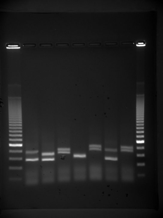
Because nucleic acids are negatively charged ions at neutral or basic pH in an aqueous environment, they can be mobilized by an electric field. Gel electrophoresis is a technique used to separate molecules on the basis of size, using this charge. The nucleic acids can be separated as whole chromosomes or fragments. The nucleic acids are loaded into a slot near the negative electrode of a semisolid, porous gel matrix and pulled toward the positive electrode at the opposite end of the gel. Smaller molecules move through the pores in the gel faster than larger molecules; this difference in the rate of migration separates the fragments on the basis of size. There are molecular weight standard samples that can be run alongside the molecules to provide a size comparison. Nucleic acids in a gel matrix can be observed using various fluorescent or colored dyes. Distinct nucleic acid fragments appear as bands at specific distances from the top of the gel (the negative electrode end) on the basis of their size ( [link] ). A mixture of genomic DNA fragments of varying sizes appear as a long smear, whereas uncut genomic DNA is usually too large to run through the gel and forms a single large band at the top of the gel.

Photo shows an agarose gel illuminated under UV light. The gel contains nine lanes from left to right. Each lane was loaded with a sample containing DNA fragments of differing size that separated as they travelled through the gel from top to bottom. The DNA appears as thin, white bands on a black background. Lanes one and nine contain many bands from a DNA standard. These bands are closely spaced toward the top, and spaced farther apart further down the gel. Lanes two through eight contain one or two bands each. Some of these bands are identical in size and run the same distance into the gel. Others run a slightly different distance, indicating a small difference in size.
Shown are DNA fragments from seven samples run on a gel, stained with a fluorescent dye, and viewed under UV light. (credit: James Jacob, Tompkins Cortland Community College)

Amplification of nucleic acid fragments by polymerase chain reaction

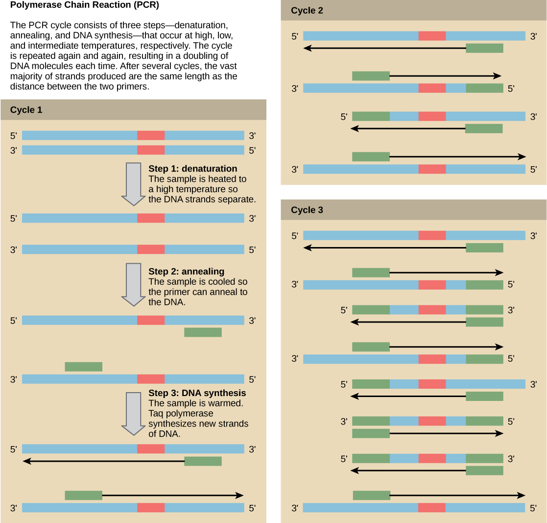
Although genomic DNA is visible to the naked eye when it is extracted in bulk, DNA analysis often requires focusing on one or more specific regions of the genome. Polymerase chain reaction ( PCR ) is a technique used to amplify specific regions of DNA for further analysis ( [link] ). PCR is used for many purposes in laboratories, such as the cloning of gene fragments to analyze genetic diseases, identification of contaminant foreign DNA in a sample, and the amplification of DNA for sequencing. More practical applications include the determination of paternity and detection of genetic diseases.

Illustration shows the amplification of a DNA sequence by the polymerase chain reaction. PCR consists of three steps—denaturation, annealing, and DNA synthesis—that occur at high, low, and intermediate temperatures. In step 1, the denaturation step, the sample is heated to a high temperature so the DNA strands separate. In step 2, annealing, the sample is cooled so two primers can anneal to the two strands of DNA. The primers are spaced such that the sequence of interest between them will be amplified. In step 3, DNA synthesis, the sample is warmed to the optimal temperature for Taq polymerase, which synthesizes the complementary strand from the primer to the 3' end of the molecule. This cycle is repeated again and again. Each time, the newly synthesized strands serve as templates so that the amount of DNA doubles with each cycle. As the cycles continue, more and more strands are the size of the distance between the two primers; in the end, the vast majority of strands are this size.
Polymerase chain reaction, or PCR, is used to amplify a specific sequence of DNA. Primers—short pieces of DNA complementary to each end of the target sequence—are combined with genomic DNA, Taq polymerase, and deoxynucleotides. Taq polymerase is a DNA polymerase isolated from the thermostable bacterium Thermus aquaticus that is able to withstand the high temperatures used in PCR. Thermus aquaticus grows in the Lower Geyser Basin of Yellowstone National Park. Reverse transcriptase PCR (RT-PCR) is similar to PCR, but cDNA is made from an RNA template before PCR begins.

Questions & Answers

what is biology
Hajah Reply
the study of living organisms and their interactions with one another and their environments
AI-Robot
what is biology
Victoria Reply
HOW CAN MAN ORGAN FUNCTION
Alfred Reply
the diagram of the digestive system
Assiatu Reply
allimentary cannel
Ogenrwot
How does twins formed
William Reply
They formed in two ways first when one sperm and one egg are splited by mitosis or two sperm and two eggs join together
Oluwatobi
what is genetics
Josephine Reply
Genetics is the study of heredity
Misack
how does twins formed?
Misack
What is manual
Hassan Reply
discuss biological phenomenon and provide pieces of evidence to show that it was responsible for the formation of eukaryotic organelles
Joseph Reply
what is biology
Yousuf Reply
the study of living organisms and their interactions with one another and their environment.
Wine
discuss the biological phenomenon and provide pieces of evidence to show that it was responsible for the formation of eukaryotic organelles in an essay form
Joseph Reply
what is the blood cells
Shaker Reply
list any five characteristics of the blood cells
Shaker
lack electricity and its more savely than electronic microscope because its naturally by using of light
Abdullahi Reply
advantage of electronic microscope is easily and clearly while disadvantage is dangerous because its electronic. advantage of light microscope is savely and naturally by sun while disadvantage is not easily,means its not sharp and not clear
Abdullahi
cell theory state that every organisms composed of one or more cell,cell is the basic unit of life
Abdullahi
is like gone fail us
DENG
cells is the basic structure and functions of all living things
Ramadan
What is classification
ISCONT Reply
is organisms that are similar into groups called tara
Yamosa
in what situation (s) would be the use of a scanning electron microscope be ideal and why?
Kenna Reply
A scanning electron microscope (SEM) is ideal for situations requiring high-resolution imaging of surfaces. It is commonly used in materials science, biology, and geology to examine the topography and composition of samples at a nanoscale level. SEM is particularly useful for studying fine details,
Hilary
cell is the building block of life.
Condoleezza Reply
Got questions? Join the online conversation and get instant answers!
Jobilize.com Reply

Get Jobilize Job Search Mobile App in your pocket Now!

Get it on Google Play Download on the App Store Now


Source:  OpenStax, Genetics and evolution. OpenStax CNX. Aug 07, 2014 Download for free at https://legacy.cnx.org/content/col11595/1.2
Google Play and the Google Play logo are trademarks of Google Inc.

Notification Switch

Would you like to follow the 'Genetics and evolution' conversation and receive update notifications?

Ask