<< Chapter < Page Chapter >> Page >

Deriving moles from grams for a compound

Our bodies synthesize protein from amino acids. One of these amino acids is glycine, which has the molecular formula C 2 H 5 O 2 N. How many moles of glycine molecules are contained in 28.35 g of glycine?

Solution

We can derive the number of moles of a compound from its mass following the same procedure we used for an element in [link] :

A diagram of two boxes connected by a right-facing arrow is shown. The box on the left contains the phrase, “Mass of C subscript 2 H subscript 5 O subscript 2 N ( g )” while the box on the right contains the phrase, “Moles of C subscript 2 H subscript 5 O subscript 2 N ( mol ).” There is a phrase under the arrow that says “Divide by molar mass (g / mol).”

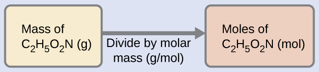
The molar mass of glycine is required for this calculation, and it is computed in the same fashion as its molecular mass. One mole of glycine, C 2 H 5 O 2 N, contains 2 moles of carbon, 5 moles of hydrogen, 2 moles of oxygen, and 1 mole of nitrogen:

A table is shown that is made up of six columns and six rows. The header row reads: “Element,” “Quantity (mol element / mol compound,” a blank space, “Molar mass (g / mol element),” a blank space, and “Subtotal (a m u).” The first column contains the symbols “C,” “H,” “O,” “N,” and a merged cell. The merged cell runs the width of the first five columns. The second column contains the numbers “2,” “5,” “2,” and “1” as well as the merged cell. The third column contains the multiplication symbol in each cell except for the last, merged cell. The fourth column contains the numbers “12.01,” “1.008,” “16.00,” and “14.007” as well as the merged cell. The fifth column contains the symbol “=” in each cell except for the last, merged cell. The sixth column contains the values “24.02,” “5.040,” “32.00,” “14.007,” and “75.07.” There is a thick black line under the number 14.007. The merged cell under the first five columns reads “Molar mass (g / mol compound). There is a ball-and-stick drawing to the right of this table. It shows a black sphere that forms a double bond with a slightly smaller red sphere, a single bond with another red sphere, and a single bond with another black sphere. The red sphere that forms a single bond with the black sphere also forms a single bond with a smaller, white sphere. The second black sphere forms a single bond with a smaller, white sphere and a smaller blue sphere. The blue sphere forms a single bond with two smaller, white spheres each.

The provided mass of glycine (~28 g) is a bit more than one-third the molar mass (~75 g/mol), so we would expect the computed result to be a bit greater than one-third of a mole (~0.33 mol). Dividing the compound’s mass by its molar mass yields:

28.35 g glycine ( mol glycine 75.07 g ) = 0.378 mol glycine

This result is consistent with our rough estimate.

Check your learning

How many moles of sucrose, C 12 H 22 O 11 , are in a 25-g sample of sucrose?

Answer:

0.073 mol

Got questions? Get instant answers now!

Deriving grams from moles for a compound

Vitamin C is a covalent compound with the molecular formula C 6 H 8 O 6 . The recommended daily dietary allowance of vitamin C for children aged 4–8 years is 1.42 × 10 −4 mol. What is the mass of this allowance in grams?

Solution

As for elements, the mass of a compound can be derived from its molar amount as shown:

A diagram of two boxes connected by a right-facing arrow is shown. The box on the left contains the phrase, “Moles of vitamin C ( mol )” while the one the right contains the phrase, “Mass of vitamin C ( g )”. There is a phrase under the arrow that says “Multiply by molar mass (g / mol).”

The molar mass for this compound is computed to be 176.124 g/mol. The given number of moles is a very small fraction of a mole (~10 −4 or one-ten thousandth); therefore, we would expect the corresponding mass to be about one-ten thousandth of the molar mass (~0.02 g). Performing the calculation, we get:

1.42 × 10 −4 mol vitamin C ( 176.124 g mol vitamin C ) = 0.0250 g vitamin C

This is consistent with the anticipated result.

Check your learning

What is the mass of 0.443 mol of hydrazine, N 2 H 4 ?

Answer:

14.2 g

Got questions? Get instant answers now!

Deriving the number of atoms and molecules from the mass of a compound

A packet of an artificial sweetener contains 40.0 mg of saccharin (C 7 H 5 NO 3 S), which has the structural formula:

A diagram of a molecule is shown that is made up of two ring structures attached together. The left ring is hexagonal in shape with C atoms at each point of the ring and alternating single and double bonds. A double bond occurs between the C atom at the top vertex of the hexagon and the C atom down and to the left of it. The C atoms on the left, top, and bottom of the structure form a single bond to an H atom each. The two right C atoms make up one side of a pentagon and the other points of the pentagon are made up of a C atom, an N atom, and an S atom if read clockwise. The C atom forms a double bond with an O atom. The N atom forms a single bond with an H atom. The S atom forms two double bonds to two O atoms.

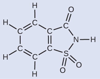
Given that saccharin has a molar mass of 183.18 g/mol, how many saccharin molecules are in a 40.0-mg (0.0400-g) sample of saccharin? How many carbon atoms are in the same sample?

Solution

The number of molecules in a given mass of compound is computed by first deriving the number of moles, as demonstrated in [link] , and then multiplying by Avogadro’s number:

A diagram of three boxes connected by a right-facing arrow in between each is shown. The box on the left contains the phrase, “Mass of C subscript seven H subscript five N O subscript three S ( g ),” the middle box reads, “Moles of C subscript seven H subscript five N O subscript three S ( mol ),” while the one on the right contains the phrase, “Number of C subscript seven H subscript five N O subscript three S molecules.” There is a phrase under the left arrow that says, “Divide by molar mass (g / mol),” and under the right arrow it states, “Multiply by Avogadro’s number ( mol superscript negative one).”

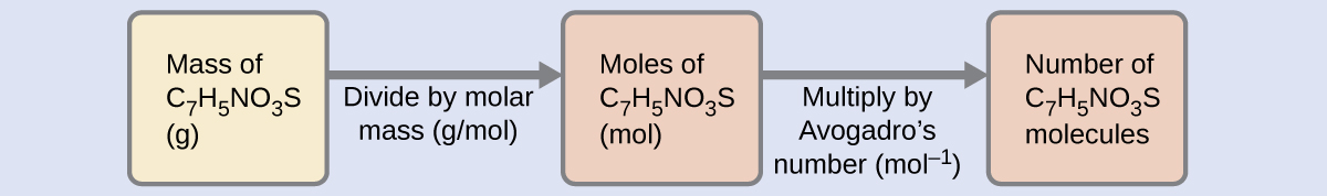
Using the provided mass and molar mass for saccharin yields:

0.0400 g C 7 H 5 NO 3 S ( mol C 7 H 5 NO 3 S 183.18 g C 7 H 5 NO 3 S ) ( 6.022 × 10 23 C 7 H 5 NO 3 S molecules 1 mol C 7 H 5 NO 3 S ) = 1.31 × 10 20 C 7 H 5 NO 3 S molecules

The compound’s formula shows that each molecule contains seven carbon atoms, and so the number of C atoms in the provided sample is:

1.31 × 10 20 C 7 H 5 NO 3 S molecules ( 7 C atoms 1 C 7 H 5 NO 3 S molecule ) = 9.20 × 10 21 C atoms

Check your learning

How many C 4 H 10 molecules are contained in 9.213 g of this compound? How many hydrogen atoms?

Answer:

9.545 × 10 22 molecules C 4 H 10 ; 9.545 × 10 23 atoms H

Got questions? Get instant answers now!

Counting neurotransmitter molecules in the brain

The brain is the control center of the central nervous system ( [link] ). It sends and receives signals to and from muscles and other internal organs to monitor and control their functions; it processes stimuli detected by sensory organs to guide interactions with the external world; and it houses the complex physiological processes that give rise to our intellect and emotions. The broad field of neuroscience spans all aspects of the structure and function of the central nervous system, including research on the anatomy and physiology of the brain. Great progress has been made in brain research over the past few decades, and the BRAIN Initiative, a federal initiative announced in 2013, aims to accelerate and capitalize on these advances through the concerted efforts of various industrial, academic, and government agencies (more details available at www.whitehouse.gov/share/brain-initiative).

Two pictures are shown. The left picture shows the human brain. The right picture is a microscopic image that depicts two large irregularly shaped masses in a field of threadlike material interspersed with smaller, relatively round masses. The two larger masses are labeled with arrows and the phrase “Neuron cells.”
(a) A typical human brain weighs about 1.5 kg and occupies a volume of roughly 1.1 L. (b) Information is transmitted in brain tissue and throughout the central nervous system by specialized cells called neurons (micrograph shows cells at 1600× magnification).

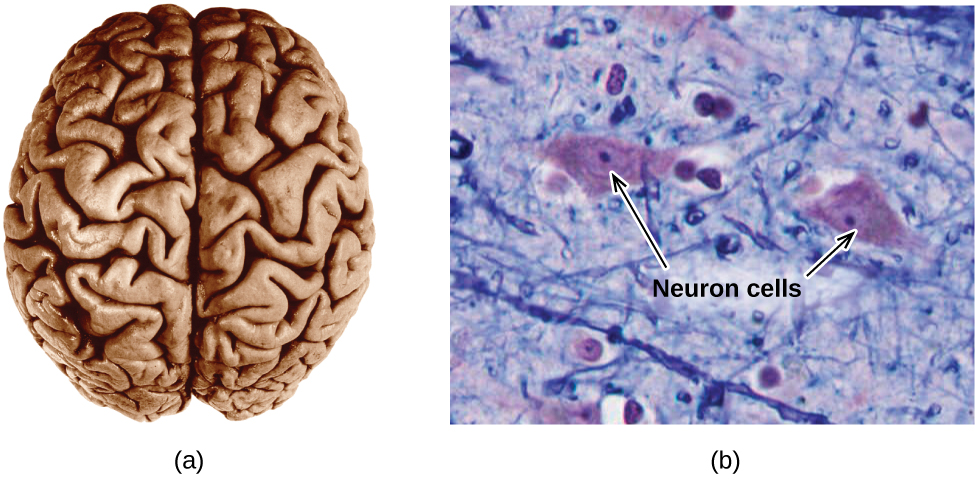
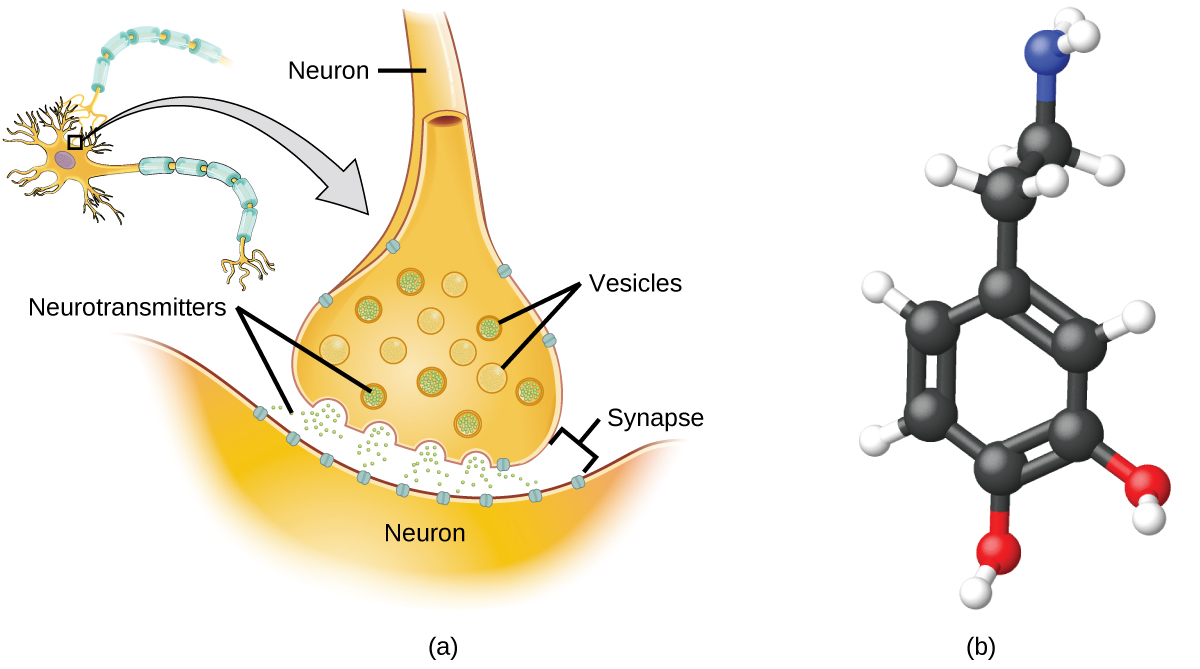
Specialized cells called neurons transmit information between different parts of the central nervous system by way of electrical and chemical signals. Chemical signaling occurs at the interface between different neurons when one of the cells releases molecules (called neurotransmitters) that diffuse across the small gap between the cells (called the synapse) and bind to the surface of the other cell. These neurotransmitter molecules are stored in small intracellular structures called vesicles that fuse to the cell wall and then break open to release their contents when the neuron is appropriately stimulated. This process is called exocytosis (see [link] ). One neurotransmitter that has been very extensively studied is dopamine, C 8 H 11 NO 2 . Dopamine is involved in various neurological processes that impact a wide variety of human behaviors. Dysfunctions in the dopamine systems of the brain underlie serious neurological diseases such as Parkinson’s and schizophrenia.

Two diagrams are shown. In the upper left corner of the left diagram, an oval with a darkened center that has five short, branching appendages and one long tail-like appendage is shown and connected by an arrow to another image. This image depicts a close-up view of the oval section and its interaction with the tail-like portion of a similar structure. The close up view is composed of a narrow tube labeled “neuron” leading down to a bulbous base that holds thirteen circles filled with small dots. These circles are labeled “vesicles.” The base of the bulbous structure is next to a curved object labeled “neuron” and very small dots are emerging from the bulb’s base and flowing toward the curved structure. The gap in between the two structures is labeled “synapse,” and the small dots are labeled “neurotransmitters.” The diagram on the right depicts a molecule composed of six black spheres connected by alternating double and single bonds in a hexagonal ring with other spheres attached to it. Three of the black spheres are connected to one smaller, white sphere each. Two of the black balls are connected to a smaller red sphere each. Each red sphere is connected to a smaller, white sphere. One black sphere is connected to another black sphere. It is connected to two smaller, white spheres and another black sphere. This second black sphere is connected to two smaller white spheres, and a slightly smaller blue sphere. The blue sphere is connected to two smaller, white spheres.
(a) Chemical signals are transmitted from neurons to other cells by the release of neurotransmitter molecules into the small gaps (synapses) between the cells. (b) Dopamine, C 8 H 11 NO 2 , is a neurotransmitter involved in a number of neurological processes.

One important aspect of the complex processes related to dopamine signaling is the number of neurotransmitter molecules released during exocytosis. Since this number is a central factor in determining neurological response (and subsequent human thought and action), it is important to know how this number changes with certain controlled stimulations, such as the administration of drugs. It is also important to understand the mechanism responsible for any changes in the number of neurotransmitter molecules released—for example, some dysfunction in exocytosis, a change in the number of vesicles in the neuron, or a change in the number of neurotransmitter molecules in each vesicle.

Significant progress has been made recently in directly measuring the number of dopamine molecules stored in individual vesicles and the amount actually released when the vesicle undergoes exocytosis. Using miniaturized probes that can selectively detect dopamine molecules in very small amounts, scientists have determined that the vesicles of a certain type of mouse brain neuron contain an average of 30,000 dopamine molecules per vesicle (about 5 × 10 −20 mol or 50 zmol). Analysis of these neurons from mice subjected to various drug therapies shows significant changes in the average number of dopamine molecules contained in individual vesicles, increasing or decreasing by up to three-fold, depending on the specific drug used. These studies also indicate that not all of the dopamine in a given vesicle is released during exocytosis, suggesting that it may be possible to regulate the fraction released using pharmaceutical therapies. Omiatek, Donna M., Amanda J. Bressler, Ann-Sofie Cans, Anne M. Andrews, Michael L. Heien, and Andrew G. Ewing. “The Real Catecholamine Content of Secretory Vesicles in the CNS Revealed by Electrochemical Cytometry.” Scientific Report 3 (2013): 1447, accessed January 14, 2015, doi:10.1038/srep01447.

Practice Key Terms 4

Get Jobilize Job Search Mobile App in your pocket Now!

Get it on Google Play Download on the App Store Now


Source:  OpenStax, Chemistry. OpenStax CNX. May 20, 2015 Download for free at http://legacy.cnx.org/content/col11760/1.9
Google Play and the Google Play logo are trademarks of Google Inc.

Notification Switch

Would you like to follow the 'Chemistry' conversation and receive update notifications?

Ask