<< Chapter < Page Chapter >> Page >

Measuring radiation exposure

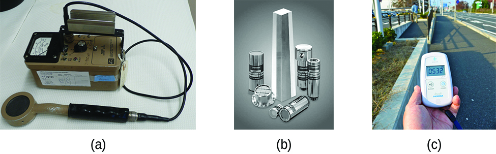
Several different devices are used to detect and measure radiation, including Geiger counters, scintillation counters (scintillators), and radiation dosimeters ( [link] ). Probably the best-known radiation instrument, the Geiger counter    (also called the Geiger-Müller counter) detects and measures radiation. Radiation causes the ionization of the gas in a Geiger-Müller tube. The rate of ionization is proportional to the amount of radiation. A scintillation counter    contains a scintillator—a material that emits light (luminesces) when excited by ionizing radiation—and a sensor that converts the light into an electric signal. Radiation dosimeters also measure ionizing radiation and are often used to determine personal radiation exposure. Commonly used types are electronic, film badge, thermoluminescent, and quartz fiber dosimeters.

Three photographs are shown and labeled “a,” “b” and “c.” Photo a shows a Geiger counter sitting on a table. It is made up of a metal box with a read-out screen and a wire leading away from the box connected to a sensor wand. Photograph b shows a collection of tall and short vertical tubes arranged in a grouping while photograph c shows a person’s hand holding a small machine with a digital readout while standing on the edge of a roadway.
Devices such as (a) Geiger counters, (b) scintillators, and (c) dosimeters can be used to measure radiation. (credit c: modification of work by “osaMu”/Wikimedia commons)

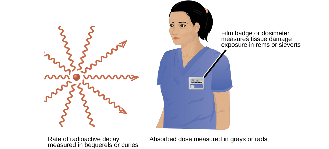
A variety of units are used to measure various aspects of radiation ( [link] ). The SI unit for rate of radioactive decay is the becquerel (Bq)    , with 1 Bq = 1 disintegration per second. The curie (Ci)    and millicurie (mCi)    are much larger units and are frequently used in medicine (1 curie = 1 Ci = 3.7 × 10 10 disintegrations per second). The SI unit for measuring radiation dose is the gray (Gy)    , with 1 Gy = 1 J of energy absorbed per kilogram of tissue. In medical applications, the radiation absorbed dose (rad)    is more often used (1 rad = 0.01 Gy; 1 rad results in the absorption of 0.01 J/kg of tissue). The SI unit measuring tissue damage caused by radiation is the sievert (Sv)    . This takes into account both the energy and the biological effects of the type of radiation involved in the radiation dose. The roentgen equivalent for man (rem) is the unit for radiation damage that is used most frequently in medicine (1 rem = 1 Sv). Note that the tissue damage units (rem or Sv) includes the energy of the radiation dose (rad or Gy) along with a biological factor referred to as the RBE (for relative biological effectiveness ) that is an approximate measure of the relative damage done by the radiation. These are related by:

number of rems = RBE × number of rads

with RBE approximately 10 for α radiation, 2(+) for protons and neutrons, and 1 for β and γ radiation.

Two images are shown. The first, labeled “Rate of radioactive decay measured in becquerels or curies,” shows a red sphere with ten red squiggly arrows facing away from it in a 360 degree circle. The second image shows the head and torso of a woman wearing medical scrubs with a badge on her chest. The caption to the badge reads “Film badge or dosimeter measures tissue damage exposure in rems or sieverts” while a phrase under this image states “Absorbed dose measured in grays or rads.”
Different units are used to measure the rate of emission from a radioactive source, the energy that is absorbed from the source, and the amount of damage the absorbed radiation does.

Units of radiation measurement

[link] summarizes the units used for measuring radiation.

Units Used for Measuring Radiation
Measurement Purpose Unit Quantity Measured Description
activity of source becquerel (Bq) radioactive decays or emissions amount of sample that undergoes 1 decay/second
curie (Ci) amount of sample that undergoes 3.7 × 10 10 decays/second
absorbed dose gray (Gy) energy absorbed per kg of tissue 1 Gy = 1 J/kg tissue
radiation absorbed dose (rad) 1 rad = 0.01 J/kg tissue
biologically effective dose sievert (Sv) tissue damage Sv = RBE × Gy
roentgen equivalent for man (rem) Rem = RBE × rad

Questions & Answers

what is biology
Hajah Reply
the study of living organisms and their interactions with one another and their environments
AI-Robot
what is biology
Victoria Reply
HOW CAN MAN ORGAN FUNCTION
Alfred Reply
the diagram of the digestive system
Assiatu Reply
allimentary cannel
Ogenrwot
How does twins formed
William Reply
They formed in two ways first when one sperm and one egg are splited by mitosis or two sperm and two eggs join together
Oluwatobi
what is genetics
Josephine Reply
Genetics is the study of heredity
Misack
how does twins formed?
Misack
What is manual
Hassan Reply
discuss biological phenomenon and provide pieces of evidence to show that it was responsible for the formation of eukaryotic organelles
Joseph Reply
what is biology
Yousuf Reply
the study of living organisms and their interactions with one another and their environment.
Wine
discuss the biological phenomenon and provide pieces of evidence to show that it was responsible for the formation of eukaryotic organelles in an essay form
Joseph Reply
what is the blood cells
Shaker Reply
list any five characteristics of the blood cells
Shaker
lack electricity and its more savely than electronic microscope because its naturally by using of light
Abdullahi Reply
advantage of electronic microscope is easily and clearly while disadvantage is dangerous because its electronic. advantage of light microscope is savely and naturally by sun while disadvantage is not easily,means its not sharp and not clear
Abdullahi
cell theory state that every organisms composed of one or more cell,cell is the basic unit of life
Abdullahi
is like gone fail us
DENG
cells is the basic structure and functions of all living things
Ramadan
What is classification
ISCONT Reply
is organisms that are similar into groups called tara
Yamosa
in what situation (s) would be the use of a scanning electron microscope be ideal and why?
Kenna Reply
A scanning electron microscope (SEM) is ideal for situations requiring high-resolution imaging of surfaces. It is commonly used in materials science, biology, and geology to examine the topography and composition of samples at a nanoscale level. SEM is particularly useful for studying fine details,
Hilary
cell is the building block of life.
Condoleezza Reply
Got questions? Join the online conversation and get instant answers!
Jobilize.com Reply

Get Jobilize Job Search Mobile App in your pocket Now!

Get it on Google Play Download on the App Store Now


Source:  OpenStax, Chemistry. OpenStax CNX. May 20, 2015 Download for free at http://legacy.cnx.org/content/col11760/1.9
Google Play and the Google Play logo are trademarks of Google Inc.

Notification Switch

Would you like to follow the 'Chemistry' conversation and receive update notifications?

Ask