

| << Chapter < Page | Chapter >> Page > |
Cytokinesis, the physical separation of the cytoplasmic components into two daughter cells, occurs without reformation of the nuclei in other organisms. In nearly all animals, cytokinesis separates the cell contents by a cleavage furrow. At each pole, there is just one member of each pair of the homologous chromosomes, so only one full set of the chromosomes is present. This is why the cells are considered haploid—there is only one chromosome set, even though there are duplicate copies of the set because each homolog still consists of two sister chromatids that are still attached to each other. However, although the sister chromatids were once duplicates of the same chromosome, they are no longer identical at this stage because of crossovers.
Review the process of meiosis, observing how chromosomes align and migrate, at this site .
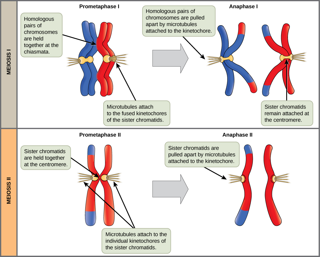
In meiosis II, the connected sister chromatids remaining in the haploid cells from meiosis I will be split to form four haploid cells. In some species, cells enter a brief interphase, or interkinesis , that lacks an S phase, before entering meiosis II. Chromosomes are not duplicated during interkinesis. The two cells produced in meiosis I go through the events of meiosis II in synchrony. Overall, meiosis II resembles the mitotic division of a haploid cell.
In prophase II, if the chromosomes decondensed in telophase I, they condense again. If nuclear envelopes were formed, they fragment into vesicles. The centrosomes duplicated during interkinesis move away from each other toward opposite poles, and new spindles are formed. In prometaphase II, the nuclear envelopes are completely broken down, and the spindle is fully formed. Each sister chromatid forms an individual kinetochore that attaches to microtubules from opposite poles. In metaphase II, the sister chromatids are maximally condensed and aligned at the center of the cell. In anaphase II, the sister chromatids are pulled apart by the spindle fibers and move toward opposite poles.
In telophase II, the chromosomes arrive at opposite poles and begin to decondense. Nuclear envelopes form around the chromosomes. Cytokinesis separates the two cells into four genetically unique haploid cells. At this point, the nuclei in the newly produced cells are both haploid and have only one copy of the single set of chromosomes. The cells produced are genetically unique because (assuming there was parental genetic variation) of the random assortment of paternal and maternal homologs and because of the recombination of maternal and paternal segments of chromosomes—with their sets of genes—that occurs during crossover.
Mitosis and meiosis, which are both forms of division of the nucleus in eukaryotic cells, share some similarities, but also exhibit distinct differences that lead to their very different outcomes. Mitosis is a single nuclear division that results in two nuclei, usually partitioned into two new cells. The nuclei resulting from a mitotic division are genetically identical to the original. They have the same number of sets of chromosomes: one in the case of haploid cells, and two in the case of diploid cells. On the other hand, meiosis is two nuclear divisions that result in four nuclei, usually partitioned into four new cells. The nuclei resulting from meiosis are never genetically identical, and they contain one chromosome set only—this is half the number of the original cell, which was diploid ( [link] ).
Notification Switch
Would you like to follow the 'Human biology' conversation and receive update notifications?

|
|

|
|

|
|
|
|

|
|
|
|
|
|
|
|

|
|

|
|

|