<< Chapter < Page Chapter >> Page >

The osteoblast    is the bone cell responsible for forming new bone and is found in the growing portions of bone, including the periosteum and endosteum. Osteoblasts, which do not divide, synthesize and secrete the collagen matrix and calcium salts. As the secreted matrix surrounding the osteoblast calcifies, the osteoblast become trapped within it; as a result, it changes in structure and becomes an osteocyte    , the primary cell of mature bone and the most common type of bone cell. Each osteocyte is located in a space called a lacuna and is surrounded by bone tissue. Osteocytes maintain the mineral concentration of the matrix via the secretion of enzymes. Like osteoblasts, osteocytes lack mitotic activity. They can communicate with each other and receive nutrients via long cytoplasmic processes that extend through canaliculi    (singular = canaliculus), channels within the bone matrix.

If osteoblasts and osteocytes are incapable of mitosis, then how are they replenished when old ones die? The answer lies in the properties of a third category of bone cells—the osteogenic cell    . These osteogenic cells are undifferentiated with high mitotic activity and they are the only bone cells that divide. Immature osteogenic cells are found in the deep layers of the periosteum and the marrow. They differentiate and develop into osteoblasts.

The dynamic nature of bone means that new tissue is constantly formed, and old, injured, or unnecessary bone is dissolved for repair or for calcium release. The cell responsible for bone resorption, or breakdown, is the osteoclast    . They are found on bone surfaces, are multinucleated, and originate from monocytes and macrophages, two types of white blood cells, not from osteogenic cells. Osteoclasts are continually breaking down old bone while osteoblasts are continually forming new bone. The ongoing balance between osteoblasts and osteoclasts is responsible for the constant but subtle reshaping of bone. [link] reviews the bone cells, their functions, and locations.

Bone Cells
Cell type Function Location
Osteogenic cells Develop into osteoblasts Deep layers of the periosteum and the marrow
Osteoblasts Bone formation Growing portions of bone, including periosteum and endosteum
Osteocytes Maintain mineral concentration of matrix Entrapped in matrix
Osteoclasts Bone resorption Bone surfaces and at sites of old, injured, or unneeded bone

Compact and spongy bone

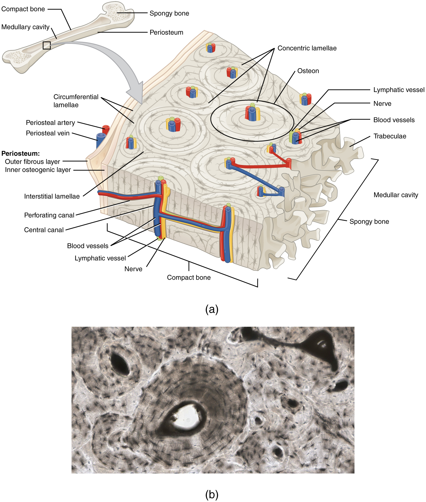
The differences between compact and spongy bone are best explored via their histology. Most bones contain compact and spongy osseous tissue, but their distribution and concentration vary based on the bone’s overall function. Compact bone is dense so that it can withstand compressive forces, while spongy (cancellous) bone has open spaces and supports shifts in weight distribution.

Compact bone

Compact bone is the denser, stronger of the two types of bone tissue ( [link] ). It can be found under the periosteum and in the diaphyses of long bones, where it provides support and protection.

Diagram of compact bone

A generic long bone is shown at the top of this illustration. The bone is split in half lengthwise to show its internal anatomy. The outer gray covering of the bone is labeled the periosteum. Within the periosteum is a thin layer of compact bone. The compact bone surrounds a central cavity called the medullary cavity. The medullary cavity is filled with spongy bone at the two epiphyses. A callout box shows that the main image is zooming in on the compact bone on the left side of the bone. On the main image, the periosteum is being peeled back to show its two layers. The outer layer of the periosteum is the outer fibrous layer. This layer has a periosteal artery and a periosteal vein running along its outside edge. The inner layer of the periosteum is labeled the inner osteogenic layer. The compact bone lies to the right of the periosteum and occupies the majority of the main image. Two flat layers of compact bone line the inner surface of the ostegenic periosteum. These sheets of compact bone are called the circumferential lamellae. The majority of the compact bone has lamellae running perpendicular to that of the circumferential lamellae. These concentric lamellae are arranged in a series of concentric tubes. There are small cavities between the layers of concentric lamellae called lacunae. The centermost concentric lamella surrounds a hollow central canal. A blue vein, a red artery, a yellow nerve and a green lymph vessel run vertically through the central canal. A set of concentric lamellae, its associated lacunae and the vessels and nerves of the central canal are collectively called an osteon. The front edge of the diagram shows a longitudinal cross section of one of the osteons. The vessels and nerve are visible running through the center of the osteon throughout its length. In addition, blood vessels can run from the periosteum through the sides of the osteons and connect with the vessels of the central canal. The blood vessels travel through the sides of the osteons via a perforating canal. The open areas between neighboring osteons are also filled with compact bone. This “filler” bone is referred to as the interstitial lamellae. At the far right of the compact bone, the edge of the spongy bone is visible. The spongy bone is a series of crisscrossing bony arches called trabeculae. There are many open spaces between the trabeculae, giving the spongy bone its sponge-like appearance.
(a) This cross-sectional view of compact bone shows the basic structural unit, the osteon. (b) In this micrograph of the osteon, you can clearly see the concentric lamellae and central canals. LM × 40. (Micrograph provided by the Regents of University of Michigan Medical School © 2012)

Questions & Answers

Discuss the differences between taste and flavor, including how other sensory inputs contribute to our  perception of flavor.
John Reply
taste refers to your understanding of the flavor . while flavor one The other hand is refers to sort of just a blend things.
Faith
While taste primarily relies on our taste buds, flavor involves a complex interplay between taste and aroma
Kamara
which drugs can we use for ulcers
Ummi Reply
omeprazole
Kamara
what
Renee
what is this
Renee
is a drug
Kamara
of anti-ulcer
Kamara
Omeprazole Cimetidine / Tagament For the complicated once ulcer - kit
Patrick
what is the function of lymphatic system
Nency Reply
Not really sure
Eli
to drain extracellular fluid all over the body.
asegid
The lymphatic system plays several crucial roles in the human body, functioning as a key component of the immune system and contributing to the maintenance of fluid balance. Its main functions include: 1. Immune Response: The lymphatic system produces and transports lymphocytes, which are a type of
asegid
to transport fluids fats proteins and lymphocytes to the blood stream as lymph
Adama
what is anatomy
Oyindarmola Reply
Anatomy is the identification and description of the structures of living things
Kamara
what's the difference between anatomy and physiology
Oyerinde Reply
Anatomy is the study of the structure of the body, while physiology is the study of the function of the body. Anatomy looks at the body's organs and systems, while physiology looks at how those organs and systems work together to keep the body functioning.
AI-Robot
what is enzymes all about?
Mohammed Reply
Enzymes are proteins that help speed up chemical reactions in our bodies. Enzymes are essential for digestion, liver function and much more. Too much or too little of a certain enzyme can cause health problems
Kamara
yes
Prince
how does the stomach protect itself from the damaging effects of HCl
Wulku Reply
little girl okay how does the stomach protect itself from the damaging effect of HCL
Wulku
it is because of the enzyme that the stomach produce that help the stomach from the damaging effect of HCL
Kamara
function of digestive system
Ali Reply
function of digestive
Ali
the diagram of the lungs
Adaeze Reply
what is the normal body temperature
Diya Reply
37 degrees selcius
Xolo
37°c
Stephanie
please why 37 degree selcius normal temperature
Mark
36.5
Simon
37°c
Iyogho
the normal temperature is 37°c or 98.6 °Fahrenheit is important for maintaining the homeostasis in the body the body regular this temperature through the process called thermoregulation which involves brain skin muscle and other organ working together to maintain stable internal temperature
Stephanie
37A c
Wulku
what is anaemia
Diya Reply
anaemia is the decrease in RBC count hemoglobin count and PVC count
Eniola
what is the pH of the vagina
Diya Reply
how does Lysin attack pathogens
Diya
acid
Mary
I information on anatomy position and digestive system and there enzyme
Elisha Reply
anatomy of the female external genitalia
Muhammad Reply
Organ Systems Of The Human Body (Continued) Organ Systems Of The Human Body (Continued)
Theophilus Reply
what's lochia albra
Kizito

Get Jobilize Job Search Mobile App in your pocket Now!

Get it on Google Play Download on the App Store Now


Source:  OpenStax, Anatomy & Physiology. OpenStax CNX. Feb 04, 2016 Download for free at http://legacy.cnx.org/content/col11496/1.8
Google Play and the Google Play logo are trademarks of Google Inc.

Notification Switch

Would you like to follow the 'Anatomy & Physiology' conversation and receive update notifications?

Ask