<< Chapter < Page Chapter >> Page >
By the end of this section, you will be able to:
  • Identify the anatomical features of a bone
  • Describe the histology of bone tissue
  • Compare and contrast compact and spongy bone
  • Identify the structures that compose compact and spongy bone
  • Describe how bones are nourished and innervated

Bone tissue (osseous tissue) differs greatly from other tissues in the body. Bone is hard and many of its functions depend on that characteristic hardness. Later discussions in this chapter will show that bone is also dynamic in that its shape adjusts to accommodate stresses. This section will examine the gross anatomy of bone first and then move on to its histology.

Gross anatomy of bone

The structure of a long bone allows for the best visualization of all of the parts of a bone ( [link] ). A long bone has two parts: the diaphysis    and the epiphysis    . The diaphysis is the tubular shaft that runs between the proximal and distal ends of the bone. The hollow region in the diaphysis is called the medullary cavity    , which is filled with yellow marrow. The walls of the diaphysis are composed of dense and hard compact bone    .

Anatomy of a long bone

This illustration depicts an anterior view of the right femur, or thigh bone. The inferior end that connects to the knee is at the bottom of the diagram and the superior end that connects to the hip is at the top of the diagram. The bottom end of the bone contains a smaller lateral bulge and a larger medial bulge. A blue articular cartilage covers the inner half of each bulge as well as the small trench that runs between the bulges. This area of the inferior end of the bone is labeled the distal epiphysis. Above the distal epiphysis is the metaphysis, where the bone tapers from the wide epiphysis into the relatively thin shaft. The entire length of the shaft is the diaphysis. The superior half of the femur is cut away to show its internal contents. The bone is covered with an outer translucent sheet called the periosteum. At the midpoint of the diaphysis, a nutrient artery travels through the periosteum and into the inner layers of the bone. The periosteum surrounds a white cylinder of solid bone labeled compact bone. The cavity at the center of the compact bone is called the medullary cavity. The inner layer of the compact bone that lines the medullary cavity is called the endosteum. Within the diaphysis, the medullary cavity contains a cylinder of yellow bone marrow that is penetrated by the nutrient artery. The superior end of the femur is also connected to the diaphysis by a metaphysis. In this upper metaphysis, the bone gradually widens between the diaphysis and the proximal epiphysis. The proximal epiphysis of the femur is roughly hexagonal in shape. However, the upper right side of the hexagon has a large, protruding knob. The femur connects and rotates within the hip socket at this knob. The knob is covered with a blue colored articular cartilage. The internal anatomy of the upper metaphysis and proximal epiphysis are revealed. The medullary cavity in these regions is filled with the mesh like spongy bone. Red bone marrow occupies the many cavities within the spongy bone. There is a clear, white line separating the spongy bone of the upper metaphysis with that of the proximal epiphysis. This line is labeled the epiphyseal line.
A typical long bone shows the gross anatomical characteristics of bone.

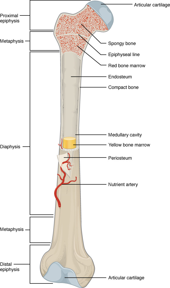
The wider section at each end of the bone is called the epiphysis (plural = epiphyses), which is filled with spongy bone. Red marrow fills the spaces in the spongy bone. Each epiphysis meets the diaphysis at the metaphysis, the narrow area that contains the epiphyseal plate    (growth plate), a layer of hyaline (transparent) cartilage in a growing bone. When the bone stops growing in early adulthood (approximately 18–21 years), the cartilage is replaced by osseous tissue and the epiphyseal plate becomes an epiphyseal line.

Bone cells and tissue

Bone contains a relatively small number of cells entrenched in a matrix of collagen fibers that provide a surface for inorganic salt crystals to adhere. These salt crystals form when calcium phosphate and calcium carbonate combine to create hydroxyapatite, which incorporates other inorganic salts like magnesium hydroxide, fluoride, and sulfate as it crystallizes, or calcifies, on the collagen fibers. The hydroxyapatite crystals give bones their hardness and strength, while the collagen fibers give them flexibility so that they are not brittle.

Although bone cells compose a small amount of the bone volume, they are crucial to the function of bones. Four types of cells are found within bone tissue: osteoblasts, osteocytes, osteogenic cells, and osteoclasts ( [link] ).

Bone cells

The top of this diagram shows the cross section of a generic bone with three zoom in boxes. The first box is on the periosteum. The second box is on the middle of the compact bone layer. The third box is on the inner edge of the compact bone where it transitions into the spongy bone. The callout in the periosteum points to two images. In the first image, four osteoblast cells are sitting end to end on the periosteum. The osteoblasts are roughly square shaped, except for one of the cells which is developing small, finger like projections. The caption says, “Osteoblasts form the matrix of the bone.” The second image called out from the periosteum shows a large, amorphous osteogenic cell sitting on the periosteum. The osteogenic cell is surrounded on both sides by a row of much smaller osteoblasts. The cell is shaped like a mushroom cap and also has finger like projections. The cell is a stem cell that develops into other bone cells. The box in the middle of the compact bone layer is pointing to an osteocyte. The osteocyte is a thin cell, roughly diamond shaped, with many branching, finger-like projections. The osteoctyes maintain bone tissue. The box at the inner edge of the compact bone is pointing to an osteoclast. The osteoclast is a large, round cell with multiple nuclei. It also has rows of fine finger like projections on its lower surface where it is sitting on the compact bone. The osteoclast reabsorbs bone.
Four types of cells are found within bone tissue. Osteogenic cells are undifferentiated and develop into osteoblasts. When osteoblasts get trapped within the calcified matrix, their structure and function changes, and they become osteocytes. Osteoclasts develop from monocytes and macrophages and differ in appearance from other bone cells.

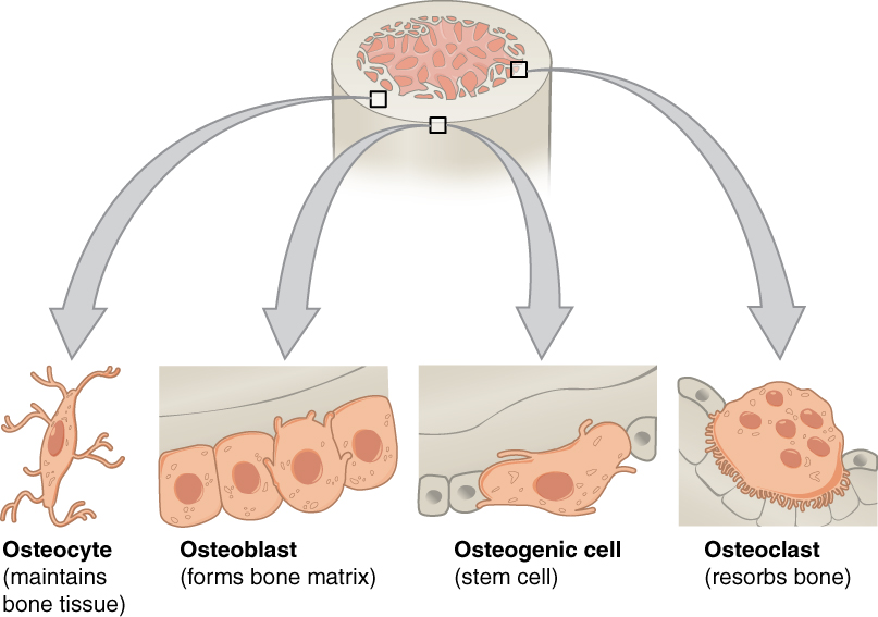
The osteoblast    is the bone cell responsible for forming new bone and is found in the growing portions of bone, including the periosteum and endosteum. Osteoblasts, which do not divide, synthesize and secrete the collagen matrix and calcium salts. As the secreted matrix surrounding the osteoblast calcifies, the osteoblast become trapped within it; as a result, it changes in structure and becomes an osteocyte    , the primary cell of mature bone and the most common type of bone cell. Each osteocyte is located in a space called a lacuna and is surrounded by bone tissue. Osteocytes maintain the mineral concentration of the matrix via the secretion of enzymes. Like osteoblasts, osteocytes lack mitotic activity. They can communicate with each other and receive nutrients via long cytoplasmic processes that extend through canaliculi    (singular = canaliculus), channels within the bone matrix.

Questions & Answers

prostaglandin and fever
Maha Reply
Discuss the differences between taste and flavor, including how other sensory inputs contribute to our  perception of flavor.
John Reply
taste refers to your understanding of the flavor . while flavor one The other hand is refers to sort of just a blend things.
Faith
While taste primarily relies on our taste buds, flavor involves a complex interplay between taste and aroma
Kamara
which drugs can we use for ulcers
Ummi Reply
omeprazole
Kamara
what
Renee
what is this
Renee
is a drug
Kamara
of anti-ulcer
Kamara
Omeprazole Cimetidine / Tagament For the complicated once ulcer - kit
Patrick
what is the function of lymphatic system
Nency Reply
Not really sure
Eli
to drain extracellular fluid all over the body.
asegid
The lymphatic system plays several crucial roles in the human body, functioning as a key component of the immune system and contributing to the maintenance of fluid balance. Its main functions include: 1. Immune Response: The lymphatic system produces and transports lymphocytes, which are a type of
asegid
to transport fluids fats proteins and lymphocytes to the blood stream as lymph
Adama
what is anatomy
Oyindarmola Reply
Anatomy is the identification and description of the structures of living things
Kamara
what's the difference between anatomy and physiology
Oyerinde Reply
Anatomy is the study of the structure of the body, while physiology is the study of the function of the body. Anatomy looks at the body's organs and systems, while physiology looks at how those organs and systems work together to keep the body functioning.
AI-Robot
what is enzymes all about?
Mohammed Reply
Enzymes are proteins that help speed up chemical reactions in our bodies. Enzymes are essential for digestion, liver function and much more. Too much or too little of a certain enzyme can cause health problems
Kamara
yes
Prince
how does the stomach protect itself from the damaging effects of HCl
Wulku Reply
little girl okay how does the stomach protect itself from the damaging effect of HCL
Wulku
it is because of the enzyme that the stomach produce that help the stomach from the damaging effect of HCL
Kamara
function of digestive system
Ali Reply
function of digestive
Ali
the diagram of the lungs
Adaeze Reply
what is the normal body temperature
Diya Reply
37 degrees selcius
Xolo
37°c
Stephanie
please why 37 degree selcius normal temperature
Mark
36.5
Simon
37°c
Iyogho
the normal temperature is 37°c or 98.6 °Fahrenheit is important for maintaining the homeostasis in the body the body regular this temperature through the process called thermoregulation which involves brain skin muscle and other organ working together to maintain stable internal temperature
Stephanie
37A c
Wulku
what is anaemia
Diya Reply
anaemia is the decrease in RBC count hemoglobin count and PVC count
Eniola
what is the pH of the vagina
Diya Reply
how does Lysin attack pathogens
Diya
acid
Mary
I information on anatomy position and digestive system and there enzyme
Elisha Reply
anatomy of the female external genitalia
Muhammad Reply
Got questions? Join the online conversation and get instant answers!
Jobilize.com Reply

Get Jobilize Job Search Mobile App in your pocket Now!

Get it on Google Play Download on the App Store Now


Source:  OpenStax, Human biology. OpenStax CNX. Dec 01, 2015 Download for free at http://legacy.cnx.org/content/col11903/1.3
Google Play and the Google Play logo are trademarks of Google Inc.

Notification Switch

Would you like to follow the 'Human biology' conversation and receive update notifications?

Ask