<< Chapter < Page Chapter >> Page >

Gametogenesis (spermatogenesis and oogenesis)

Gametogenesis, the production of sperm and eggs, involves the process of meiosis. During meiosis, two nuclear divisions separate the paired chromosomes in the nucleus and then separate the chromatids that were made during an earlier stage of the cell’s life cycle. Meiosis and its associated cell divisions produces haploid cells with half of each pair of chromosomes normally found in diploid cells. The production of sperm is called spermatogenesis    and the production of eggs is called oogenesis    .

Spermatogenesis

Spermatogenesis occurs in the wall of the seminiferous tubules, with the most primitive cells at the periphery of the tube and the most mature sperm at the lumen of the tube ( [link] ). Immediately under the capsule of the tubule are diploid, undifferentiated cells. These stem cells, each called a spermatogonium (pl. spermatogonia), go through mitosis to produce one cell that remains as a stem cell and a second cell called a primary spermatocyte that will undergo meiosis to produce sperm.

The diploid primary spermatocyte goes through meiosis I to produce two haploid cells called secondary spermatocytes. Each secondary spermatocyte divides after meiosis II to produce two cells called spermatids. The spermatids eventually reach the lumen of the tubule and grow a flagellum, becoming sperm cells. Four sperm result from each primary spermatocyte that goes through meiosis.

 Spermatogenesis begins when the 2n spermatogonium undergoes mitosis, producing more spermatogonia. The spermatogonia undergo meiosis I, producing haploid (1n) secondary spermatocytes, and meiosis II, producing spermatids. Differentiation of the spermatids results in mature sperm.
During spermatogenesis, four sperm result from each primary spermatocyte. The process also maps onto the physical structure of the wall of the seminiferous tubule, with the spermatogonia on the outer side of the tubule, and the sperm with their developing tails extended into the lumen of the tubule.

Concept in action

Visit this site to see the process of spermatogenesis.

Oogenesis

Oogenesis occurs in the outermost layers of the ovaries. As with sperm production, oogenesis starts with a germ cell. In oogenesis, this germ cell is called an oogonium and forms during the embryological development of the individual. The oogonium undergoes mitosis to produce about one to two million oocytes by the time of birth.

Oogenesis begins when the 2n oogonium undergoes mitosis, producing a primary oocyte. The primary oocytes arrest in prophase 1 before birth. After puberty, meiosis of one oocyte per menstrual cycle continues, resulting in a 1n secondary oocyte that arrests in metaphase 2 and a polar body. Upon ovulation and sperm entry, meiosis is completed and fertilization occurs, resulting in a polar body and a fertilized egg.
The process of oogenesis occurs in the ovary’s outermost layer.

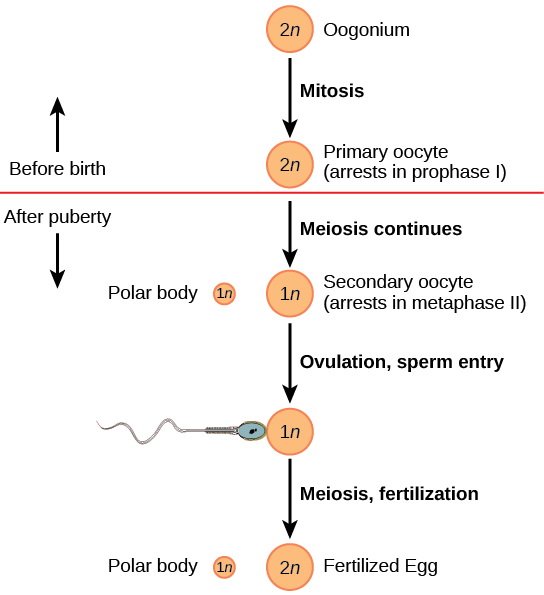
The primary oocytes begin meiosis before birth ( [link] ). However, the meiotic division is arrested in its progress in the first prophase stage. At the time of birth, all future eggs are in prophase I. This situation is in contrast with the male reproductive system in which sperm are produced continuously throughout the life of the individual. Starting at adolescence, anterior pituitary hormones cause the development of a few follicles in an ovary each month. This results in a primary oocyte finishing the first meiotic division. The cell divides unequally, with most of the cytoplasm and organelles going to one cell, called a secondary oocyte, and only one set of chromosomes and a small amount of cytoplasm going to the other cell. This second cell is called a polar body and usually dies. Cell division is again arrested, this time at metaphase II. At ovulation, this secondary oocyte is released and travels toward the uterus through the oviduct. If the secondary oocyte is fertilized, the cell continues through meiosis II, producing a second polar body and haploid egg, which fuses with the haploid sperm to form a fertilized egg (zygote) containing all 46 chromosomes.

Questions & Answers

what is phylogeny
Odigie Reply
evolutionary history and relationship of an organism or group of organisms
AI-Robot
ok
Deng
what is biology
Hajah Reply
the study of living organisms and their interactions with one another and their environments
AI-Robot
what is biology
Victoria Reply
HOW CAN MAN ORGAN FUNCTION
Alfred Reply
the diagram of the digestive system
Assiatu Reply
allimentary cannel
Ogenrwot
How does twins formed
William Reply
They formed in two ways first when one sperm and one egg are splited by mitosis or two sperm and two eggs join together
Oluwatobi
what is genetics
Josephine Reply
Genetics is the study of heredity
Misack
how does twins formed?
Misack
What is manual
Hassan Reply
discuss biological phenomenon and provide pieces of evidence to show that it was responsible for the formation of eukaryotic organelles
Joseph Reply
what is biology
Yousuf Reply
the study of living organisms and their interactions with one another and their environment.
Wine
discuss the biological phenomenon and provide pieces of evidence to show that it was responsible for the formation of eukaryotic organelles in an essay form
Joseph Reply
what is the blood cells
Shaker Reply
list any five characteristics of the blood cells
Shaker
lack electricity and its more savely than electronic microscope because its naturally by using of light
Abdullahi Reply
advantage of electronic microscope is easily and clearly while disadvantage is dangerous because its electronic. advantage of light microscope is savely and naturally by sun while disadvantage is not easily,means its not sharp and not clear
Abdullahi
cell theory state that every organisms composed of one or more cell,cell is the basic unit of life
Abdullahi
is like gone fail us
DENG
cells is the basic structure and functions of all living things
Ramadan
What is classification
ISCONT Reply
is organisms that are similar into groups called tara
Yamosa
in what situation (s) would be the use of a scanning electron microscope be ideal and why?
Kenna Reply
A scanning electron microscope (SEM) is ideal for situations requiring high-resolution imaging of surfaces. It is commonly used in materials science, biology, and geology to examine the topography and composition of samples at a nanoscale level. SEM is particularly useful for studying fine details,
Hilary
Got questions? Join the online conversation and get instant answers!
Jobilize.com Reply

Get Jobilize Job Search Mobile App in your pocket Now!

Get it on Google Play Download on the App Store Now


Source:  OpenStax, Concepts of biology. OpenStax CNX. Feb 29, 2016 Download for free at http://cnx.org/content/col11487/1.9
Google Play and the Google Play logo are trademarks of Google Inc.

Notification Switch

Would you like to follow the 'Concepts of biology' conversation and receive update notifications?

Ask