<< Chapter < Page Chapter >> Page >

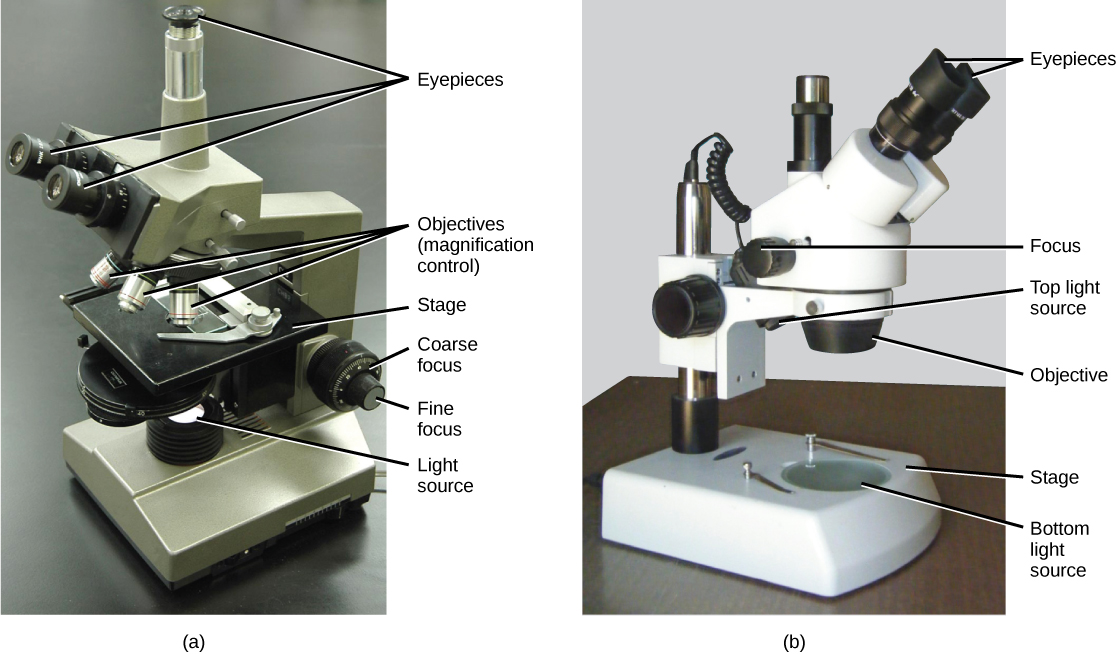
Light microscopes commonly used in the undergraduate college laboratory magnify up to approximately 400 times. Two parameters that are important in microscopy are magnification and resolving power. Magnification is the degree of enlargement of an object. Resolving power is the ability of a microscope to allow the eye to distinguish two adjacent structures as separate; the higher the resolution, the closer those two objects can be, and the better the clarity and detail of the image. When oil immersion lenses are used, magnification is usually increased to 1,000 times for the study of smaller cells, like most prokaryotic cells. Because light entering a specimen from below is focused onto the eye of an observer, the specimen can be viewed using light microscopy. For this reason, for light to pass through a specimen, the sample must be thin or translucent.

Concept in action

For another perspective on cell size, try the HowBig interactive.

A second type of microscope used in laboratories is the dissecting microscope ( [link] b ). These microscopes have a lower magnification (20 to 80 times the object size) than light microscopes and can provide a three-dimensional view of the specimen. Thick objects can be examined with many components in focus at the same time. These microscopes are designed to give a magnified and clear view of tissue structure as well as the anatomy of the whole organism. Like light microscopes, most modern dissecting microscopes are also binocular, meaning that they have two separate lens systems, one for each eye. The lens systems are separated by a certain distance, and therefore provide a sense of depth in the view of their subject to make manipulations by hand easier. Dissecting microscopes also have optics that correct the image so that it appears as if being seen by the naked eye and not as an inverted image. The light illuminating a sample under a dissecting microscope typically comes from above the sample, but may also be directed from below.

Part a: This light microscope has binocular lenses and three objective lenses. The sample stage is directly beneath the objective lens. The light microscope sits on a tabletop. Part b: The dissecting microscope has binocular eyepieces, one objective lens, and light sources from both above and below the sample stage. There is room on the stage for a three-dimensional specimen.
(a) Most light microscopes used in a college biology lab can magnify cells up to approximately 400 times. (b) Dissecting microscopes have a lower magnification than light microscopes and are used to examine larger objects, such as tissues.

Electron microscopes

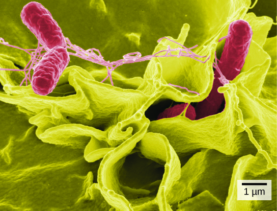
In contrast to light microscopes, electron microscopes use a beam of electrons instead of a beam of light. Not only does this allow for higher magnification and, thus, more detail ( [link] ), it also provides higher resolving power. Preparation of a specimen for viewing under an electron microscope will kill it; therefore, live cells cannot be viewed using this type of microscopy. In addition, the electron beam moves best in a vacuum, making it impossible to view living materials.

In a scanning electron microscope, a beam of electrons moves back and forth across a cell’s surface, rendering the details of cell surface characteristics by reflection. Cells and other structures are usually coated with a metal like gold. In a transmission electron microscope, the electron beam is transmitted through the cell and provides details of a cell’s internal structures. As you might imagine, electron microscopes are significantly more bulky and expensive than are light microscopes.

Part a: Salmonella viewed through a light microscope appear as tiny purple dots. Part b: In this scanning electron micrograph, the bacteria appear as three-dimensional red ovals. The human cells are much larger with a complex, folded appearance. Some of the bacteria lie on the surfaces of the human cells, and some are squeezed between them.
(a) Salmonella bacteria are viewed with a light microscope. (b) This scanning electron micrograph shows Salmonella bacteria (in red) invading human cells. (credit a: modification of work by CDC, Armed Forces Institute of Pathology, Charles N. Farmer; credit b: modification of work by Rocky Mountain Laboratories, NIAID, NIH; scale-bar data from Matt Russell)

Careers in action

Cytotechnologist

Have you ever heard of a medical test called a Pap smear ( [link] )? In this test, a doctor takes a small sample of cells from the uterine cervix of a patient and sends it to a medical lab where a cytotechnologist stains the cells and examines them for any changes that could indicate cervical cancer or a microbial infection.

Cytotechnologists ( cyto - = cell) are professionals who study cells through microscopic examinations and other laboratory tests. They are trained to determine which cellular changes are within normal limits or are abnormal. Their focus is not limited to cervical cells; they study cellular specimens that come from all organs. When they notice abnormalities, they consult a pathologist, who is a medical doctor who can make a clinical diagnosis.

Cytotechnologists play vital roles in saving people’s lives. When abnormalities are discovered early, a patient’s treatment can begin sooner, which usually increases the chances of successful treatment.

Both normal cells and cells infected with HPV have an irregular, round shape and a well-defined nucleus. The infected cells, however, are two to three times as large as uninfected cells, and some have two nuclei.
These uterine cervix cells, viewed through a light microscope, were obtained from a Pap smear. Normal cells are on the left. The cells on the right are infected with human papillomavirus. (credit: modification of work by Ed Uthman; scale-bar data from Matt Russell)

Cell theory

The microscopes we use today are far more complex than those used in the 1600s by Antony van Leeuwenhoek, a Dutch shopkeeper who had great skill in crafting lenses. Despite the limitations of his now-ancient lenses, van Leeuwenhoek observed the movements of protists (a type of single-celled organism) and sperm, which he collectively termed “animalcules.”

In a 1665 publication called Micrographia , experimental scientist Robert Hooke coined the term “cell” (from the Latin cella , meaning “small room”) for the box-like structures he observed when viewing cork tissue through a lens. In the 1670s, van Leeuwenhoek discovered bacteria and protozoa. Later advances in lenses and microscope construction enabled other scientists to see different components inside cells.

By the late 1830s, botanist Matthias Schleiden and zoologist Theodor Schwann were studying tissues and proposed the unified cell theory    , which states that all living things are composed of one or more cells, that the cell is the basic unit of life, and that all new cells arise from existing cells. These principles still stand today.

Section summary

A cell is the smallest unit of life. Most cells are so small that they cannot be viewed with the naked eye. Therefore, scientists must use microscopes to study cells. Electron microscopes provide higher magnification, higher resolution, and more detail than light microscopes. The unified cell theory states that all organisms are composed of one or more cells, the cell is the basic unit of life, and new cells arise from existing cells.

Questions & Answers

how does Neisseria cause meningitis
Nyibol Reply
what is microbiologist
Muhammad Reply
what is errata
Muhammad
is the branch of biology that deals with the study of microorganisms.
Ntefuni Reply
What is microbiology
Mercy Reply
studies of microbes
Louisiaste
when we takee the specimen which lumbar,spin,
Ziyad Reply
How bacteria create energy to survive?
Muhamad Reply
Bacteria doesn't produce energy they are dependent upon their substrate in case of lack of nutrients they are able to make spores which helps them to sustain in harsh environments
_Adnan
But not all bacteria make spores, l mean Eukaryotic cells have Mitochondria which acts as powerhouse for them, since bacteria don't have it, what is the substitution for it?
Muhamad
they make spores
Louisiaste
what is sporadic nd endemic, epidemic
Aminu Reply
the significance of food webs for disease transmission
Abreham
food webs brings about an infection as an individual depends on number of diseased foods or carriers dully.
Mark
explain assimilatory nitrate reduction
Esinniobiwa Reply
Assimilatory nitrate reduction is a process that occurs in some microorganisms, such as bacteria and archaea, in which nitrate (NO3-) is reduced to nitrite (NO2-), and then further reduced to ammonia (NH3).
Elkana
This process is called assimilatory nitrate reduction because the nitrogen that is produced is incorporated in the cells of microorganisms where it can be used in the synthesis of amino acids and other nitrogen products
Elkana
Examples of thermophilic organisms
Shu Reply
Give Examples of thermophilic organisms
Shu
advantages of normal Flora to the host
Micheal Reply
Prevent foreign microbes to the host
Abubakar
they provide healthier benefits to their hosts
ayesha
They are friends to host only when Host immune system is strong and become enemies when the host immune system is weakened . very bad relationship!
Mark
what is cell
faisal Reply
cell is the smallest unit of life
Fauziya
cell is the smallest unit of life
Akanni
ok
Innocent
cell is the structural and functional unit of life
Hasan
is the fundamental units of Life
Musa
what are emergency diseases
Micheal Reply
There are nothing like emergency disease but there are some common medical emergency which can occur simultaneously like Bleeding,heart attack,Breathing difficulties,severe pain heart stock.Hope you will get my point .Have a nice day ❣️
_Adnan
define infection ,prevention and control
Innocent
I think infection prevention and control is the avoidance of all things we do that gives out break of infections and promotion of health practices that promote life
Lubega
Heyy Lubega hussein where are u from?
_Adnan
en français
Adama
which site have a normal flora
ESTHER Reply
Many sites of the body have it Skin Nasal cavity Oral cavity Gastro intestinal tract
Safaa
skin
Asiina
skin,Oral,Nasal,GIt
Sadik
How can Commensal can Bacteria change into pathogen?
Sadik
How can Commensal Bacteria change into pathogen?
Sadik
all
Tesfaye
by fussion
Asiina
what are the advantages of normal Flora to the host
Micheal
what are the ways of control and prevention of nosocomial infection in the hospital
Micheal
what is inflammation
Shelly Reply
part of a tissue or an organ being wounded or bruised.
Wilfred
what term is used to name and classify microorganisms?
Micheal Reply
Binomial nomenclature
adeolu
Got questions? Join the online conversation and get instant answers!
Jobilize.com Reply

Get Jobilize Job Search Mobile App in your pocket Now!

Get it on Google Play Download on the App Store Now


Source:  OpenStax, Concepts of biology. OpenStax CNX. Feb 29, 2016 Download for free at http://cnx.org/content/col11487/1.9
Google Play and the Google Play logo are trademarks of Google Inc.

Notification Switch

Would you like to follow the 'Concepts of biology' conversation and receive update notifications?

Ask